. 2012 Jun;113(6):2156-66.
doi: 10.1002/jcb.24089.

Ski inhibits TGF-β/phospho-Smad3 signaling and accelerates hypertrophic differentiation in chondrocytes

Affiliations

Ski inhibits TGF-β/phospho-Smad3 signaling and accelerates hypertrophic differentiation in chondrocytes

Kyung-Ok Kim et al. J Cell Biochem. 2012 Jun.

Abstract

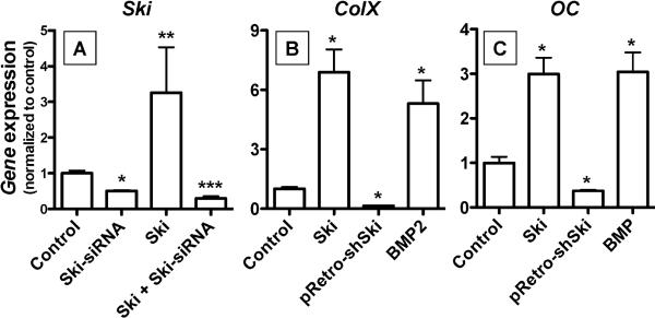
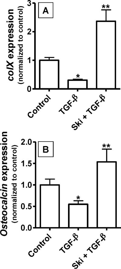
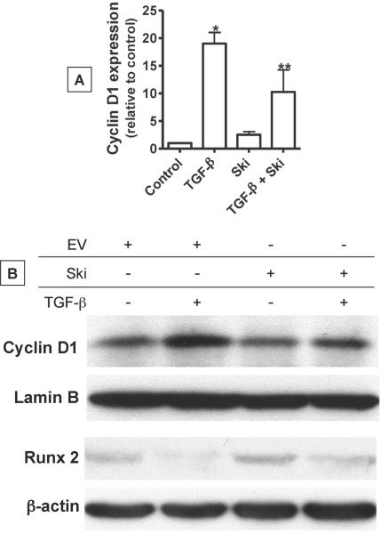
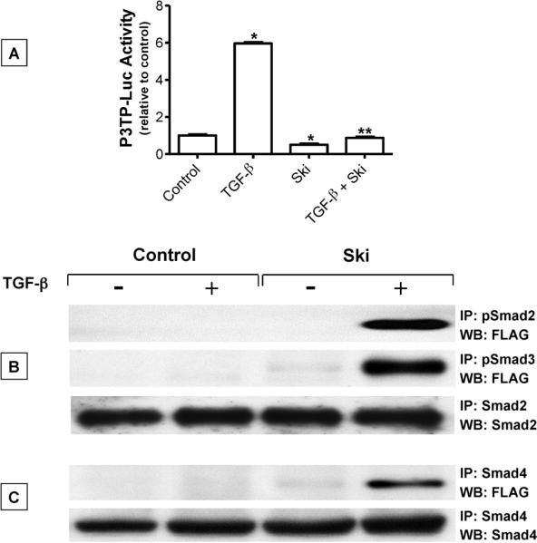
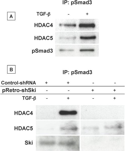
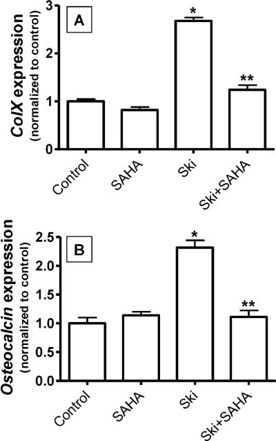
Since transforming growing factor-β (TGF-β)/Smad signaling inhibits chondrocyte maturation, endogenous negative regulators of TGF-β signaling are likely also important regulators of the chondrocyte differentiation process. One such negative regulator, Ski, is an oncoprotein that is known to inhibit TGF-β/Smad3 signaling via its interaction with phospho-Smad3 and recruitment of histone deacetylases (HDACs) to the DNA binding complex. Based on this, we hypothesized that Ski inhibits TGF-β signaling and accelerates maturation in chondrocytes via recruitment of HDACs to transcriptional complexes containing Smads. We tested this hypothesis in chick upper sternal chondrocytes (USCs), where gain and loss of Ski expression experiments were performed. Over-expression of Ski not only reversed the inhibitory effect of TGF-β on the expression of hypertrophic marker genes such as type X collagen (colX) and osteocalcin, it induced these genes basally as well. Conversely, knockdown of Ski by RNA interference led to a reduction of colX and osteocalcin expression under basal conditions. Furthermore, Ski blocked TGF-β induction of cyclinD1 and caused a basal up-regulation of Runx2, consistent with the observed acceleration of hypertrophy. Regarding mechanism, not only does Ski associate with phospho-Smad2 and 3, but its association with phospho-Smad3 is required for recruitment of HDAC4 and 5. Implicating this recruitment of HDACs in the phenotypic effects of Ski in chondrocytes, the HDAC inhibitor SAHA reversed the up-regulation of colX and osteocalcin in Ski over-expressing cells. These results suggest that inhibition of TGF-β signaling by Ski, which involves its association with phospho-Smad3 and recruitment of HDAC4 and 5, leads to accelerated chondrocyte differentiation.


Figures

Figure 1
Figure 1. Ski is expressed in cartilage
Chick tissues including lung, brain, heart, liver and sternal cartilage were harvested and proteins were extracted. Fifty μg quantities of solubilized protein were loaded and run on polyacrylamide gels, transferred to nylon membranes and Western analyses were performed to detect Ski, with detection of β-actin confirming equal loading of the gel. The analysis was performed 3 times, with the representative blot shown.
Figure 2
Figure 2. Ski mRNA expression is increased in hypertrophic USCs
Chick USCs were isolated and cultured in medium containing 50 μg/mL ascorbic acid for between 1 and 6 days. mRNA was harvested and qPCR was performed to detect the hypertrophy-associated genes colX(A) and Runx2(B) as well as Ski (C). Statistically significant differences were identified via ANOVA (N=3, p<0.05) with asterisks (*) denoting significance from the day 1 value for each gene assessed.
Figure 3
Figure 3. Gain and loss of Ski expression affects USC differentiation
Chick USCs were isolated and cultured in medium containing 50 μg/mL ascorbic acid. To confirm that experimental strategies for gain and loss of Ski expression were effective, USCs were infected with pRetro-shSkiretrovirus, transfected with cmv-Ski (Ski), treated with both reagents, or infected with nonsense shRNA virus (control). Twenty four hours post infection/transfection, mRNA was harvested and qPCR was performed to detect Ski (A). Using these Ski gain and loss of expression strategies, USCs were subsequently infected with pRetro-shSkiretrovirus, transfected with cmv-Ski or treated for 50 ng/mL BMP-2 (positive control). Control cells were infected with a nonsense shRNA retrovirus. Twenty four hours post-treatment, mRNA was harvested and qPCR was performed to quantify colX (B) and osteocalcin (C) expression levels. For all three panels, statistically significant differences were identified via ANOVA (N=3, p<0.05) with asterisks (*) denoting significance from control value for each gene assessed.
Figure 4
Figure 4. Ski reverses the inhibitory effect of TGF-β on colX and osteocalcin
As before, chick USCs were isolated and cultured in medium containing 50 μg/mL ascorbic acid. Twenty four hours after plating, cells were transfected with cmv-Ski (Ski) or an empty vector (Control). Twenty four hours post-transfection, cultures were treated with or without 5 ng/mL TGF-β. After an additional 24 hours in culture, mRNA was harvested and qPCR was performed to quantify colX (A) and osteocalcin (B) expression levels. For both panels, statistically significant differences were identified via ANOVA (N=3, p<0.05) with asterisks (*) denoting significance from the control value for each gene assessed and double asterisks (**) denoting significance from the TGF-β-treated group.
Figure 5
Figure 5. Ski stimulates Runx2 expression and reverses the effect of TGF-β on cyclin D1 and Runx2 expression
Chick USCs were isolated and cultured in medium containing 50 μg/mL ascorbic acid for 24 hours. Following this, cells were transfected with cmv-Ski (Ski) or an empty vector (Control). Twenty four hours post-transfection, cultures were treated with or without 5 ng/mL TGF-β for 24 additional hours. mRNA and total cellular protein were harvested and qPCR and Western blotting was performed to quantify cyclin D1 mRNA levels (A) and both cyclin D1 (Lamin B load control) and Runx2 (β-actin load control) protein levels (B). In panel (A), statistically significant differences were identified via ANOVA (N=3, p<0.05) with asterisks (*) denoting significance from the control value and double asterisks (**) denoting significance from the TGF-β-treated group. Blots shown in panel (B) are representative of experiments that were repeated 3 times.
Figure 6
Figure 6. Ski inhibits TGF-β signaling and co-immunoprecipitates with pSmad2, pSmad3 and Smad4
(A) Following an initial 24 hr culture period with medium containing 50 μg/mL ascorbic acid, USCs were co-transfected with P3TP-luc and SV40-renilla (transfection efficiency control) and either cmv-Ski (Ski) or an empty vector plasmid (Control). Twenty four hours later, cells were treated with or without 5 ng/mL TGF-β for 36 hours. Cell extracts were then collected and luciferase activity was measured using the Promega dual luciferase detection kit as directed by the manufacturer. Statistically significant differences were identified via ANOVA (N=3, p<0.05) with asterisks (*) denoting significance from the control group and double asterisks (**) denoting significance from the TGF-β-treated group. (B) Following an initial 24 hr culture period in medium containing 50 μg/mL ascorbic acid, USCs were transfected with cmv-Ski (Ski) or empty vector (Control). Twenty four hours later, cells were treated with or without 5 ng/mL TGF-β for 4 hours and then total cellular proteins were harvested. Immunoprecipitations were performed using either pSmad2, pSmad3, Smad2 (B) or Smad4 (C) antibody. Immunoprecipitated proteins were run out on polyacrylamide gels, transferred to nylon membranes and Western blotting was performed to detect the flag epitope (at the N-terminus of the Ski cDNA). To confirm equal loading of protein at the immunoprecipitation step, Smad2 or Smad4 immunoprecipitations were followed by detection of Smad2 (B) or Smad4 (C) respectively by Western. Blots shown in panels (B) and (C) are representative of experiments that were repeated 3 times.
Figure 7
Figure 7. Ski is required for the association between pSmad3 and HDAC4 and HDAC5
(A) Following an initial 24 hr culture period with medium containing 50 μg/mL ascorbic acid, USCs were treated with or without 5 ng/mL TGF-β for 4 hours. Total cellular proteins were harvested and immunoprecipitations were performed using pSmad3 antibody. Immunoprecipitated proteins were run out on polyacrylamide gels, transferred to nylon membranes and Western blotting was performed to detect HDAC4 and HDAC5. (B) Following initial culturing in medium containing 50 μg/mL ascorbic acid, USCs were infected with pRetro-shSki or a nonsense shRNA virus. Twenty four hours later, cells were treated with or without 5 ng/mL TGF-βfor 4 hours. Total cellular protein was harvested and immunoprecipitations were performed as described in Experimental Procedures using pSmad3 antibody. Immunoprecipitated proteins were run out on polyacrylamide gels, transferred to nylon membranes and Western blotting was performed to detect HDAC4 and HDAC5. Blots shown in panels (A) and (B) are representative of results obtained from experiments that were repeated 3 times.
Figure 8
Figure 8. Induction of colX and osteocalcin expression by Ski is blocked by HDAC inhibition
Chick USCs were isolated and cultured in medium containing 50 μg/mL ascorbic acid. Twenty four hours after plating, cells were pre-treated with the HDAC inhibitor suberoylanilidehydroxamic acid (SAHA, 5μM). Following this, cells were transfected with cmv-Ski (Ski) or empty vector (Control) and maintained in culture for an additional 24 hours in the presence of SAHA. Messenger RNA was then harvested and qPCR was performed to quantify colX (A) and osteocalcin (B) expression levels. For both panels, statistically significant differences were identified via ANOVA (N=3, p<0.05) with asterisks (*) denoting significance from the control value for each gene assessed and double asterisks (**) denoting significance from the Ski-transfected group.

Similar articles

Cited by

References

    1. Akiyoshi S, Inoue H, Hanai J, Kusanagi K, Nemoto N, Miyazono K, Kawabata M. c-Ski acts as a transcriptional co-repressor in transforming growth factor-beta signaling through interaction with smads. Journal of Biological Chemistry. 1999;274:35269–35277. - PubMed
    1. Ballock RT, Heydemann A, Wakefield LM, Flanders KC, Roberts AB, Sporn MB. TGF-beta1 prevents hypertrophy of epiphyseal chondrocytes: Regulation of gene expression for cartilage matrix proteins and metalloproteases. Developmental Biology. 1993;158:414–429. - PubMed
    1. Beier F, Ali Z, Mok D, Taylor AC, Leask T, Albanese C, Pestell RG, LuValle P. TGFbeta and PTHrP control chondrocyte proliferation by activating cyclin D1 expression. Molecular Biology of the Cell. 2001;12:3852–3863. - PMC - PubMed
    1. Beier F, Lee RJ, Taylor AC, Pestell RG, LuValle P. Identification of the cyclin D1 gene as a target of activating transcription factor 2 in chondrocytes. Proc.Natl.Acad.Sci.U.S.A. 1999;96:1433–1438. - PMC - PubMed
    1. Canalis E, Economides AN, Gazzerro E. Bone morphogenetic proteins, their antagonists, and the skeleton. Endocr.Rev. 2003;24:218–235. - PubMed

Publication types

MeSH terms

LinkOut - more resources