

Neural response to emotional stimuli during experimental human endotoxemia
- PMID: 22461242
- PMCID: PMC6870425
- DOI: 10.1002/hbm.22063
Neural response to emotional stimuli during experimental human endotoxemia
Abstract
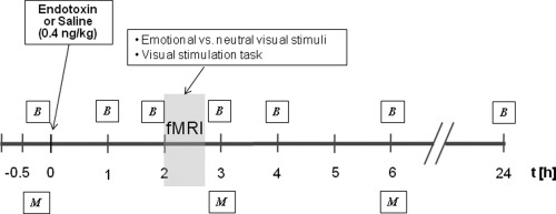
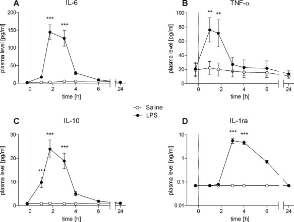
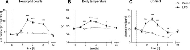
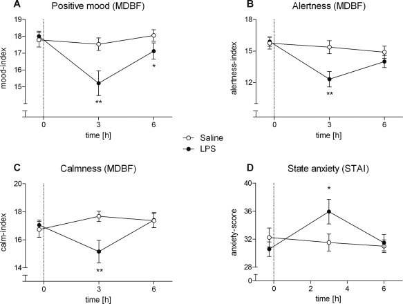
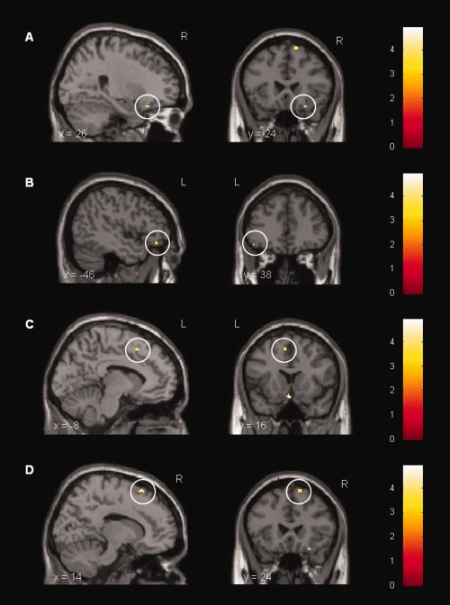
Increases in peripheral cytokines during acute inflammation may affect various neuropsychological functions. The aim of this functional magnetic resonance imaging (fMRI) study was to investigate the effects of acute endotoxemia on mood and the neural response to emotionally aversive visual stimuli in healthy human subjects. In a double-blind, randomized crossover study, 18 healthy males received a bolus injection of bacterial lipopolysaccharide (LPS; 0.4 ng/kg) or saline. Plasma levels of pro- and anti-inflammatory cytokines and cortisol as well as mood ratings were analyzed together with the blood-oxygen-level dependent (BOLD) response during the presentation of aversive versus neutral pictures. Endotoxin administration induced pronounced transient increases in plasma levels of TNF-α, IL-1ra, IL-6, IL-10, and cortisol. Positive mood was decreased and state anxiety increased. In addition, activation of right inferior orbitofrontal cortex (OFC) in response to emotional visual stimuli was significantly increased in the LPS condition. Increased prefrontal activation during the presentation of emotional material may reflect enhanced cognitive regulation of emotions as an adaptive response during an acute inflammation. These findings may have implications for the putative role of inflammatory processes in the pathophysiology of depression.
Keywords: cytokines; emotional processing; endotoxin; fMRI; peripheral inflammation; sickness behavior.
Copyright © 2012 Wiley Periodicals, Inc., a Wiley company.
Figures
References
-
- Adolphs R (2002): Neural systems for recognizing emotion. Curr Opin Neurobiol 12:169–177. - PubMed
-
- Bahador M, Cross AS (2007): From therapy to experimental model: A hundred years of endotoxin administration to human subjects. J Endotoxin Res 13:251–279. - PubMed
-
- Davidson RJ (2002): Anxiety and affective style: Role of prefrontal cortex and amygdala. Biol Psychiatry 51:68–80. - PubMed
-
- Davidson RJ, Irwin W (1999): The functional neuroanatomy of emotion and affective style. Trends Cogn Sci 3:11–21. - PubMed
Publication types
MeSH terms
Substances
LinkOut - more resources
Full Text Sources
