

Importance of myocyte-nonmyocyte interactions in cardiac development and disease
- PMID: 22461366
- PMCID: PMC3366271
- DOI: 10.1161/CIRCRESAHA.111.243899
Importance of myocyte-nonmyocyte interactions in cardiac development and disease
Abstract
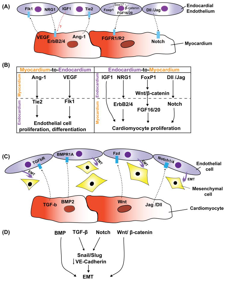
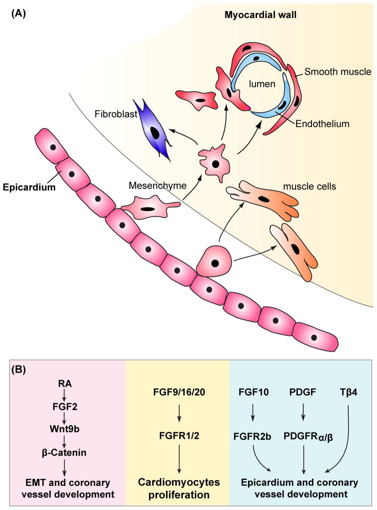
Emerging data in the field of cardiac development as well as repair and regeneration indicate a complex and important interplay between endocardial, epicardial, and myofibroblast populations that is critical for cardiomyocyte differentiation and postnatal function. For example, epicardial cells have been shown to generate cardiac myofibroblasts and may be one of the primary sources for this cell lineage during development. Moreover, paracrine signaling from the epicardium and endocardium is critical for proper development of the heart and pathways such as Wnt, fibroblast growth factor, and retinoic acid signaling have been shown to be key players in this process. Despite this progress, interactions between nonmyocyte cells and cardiomyocytes in the heart are still poorly understood. We review the various nonmyocyte-myocyte interactions that occur in the heart and how these interactions, primarily through signaling networks, help direct cardiomyocyte differentiation and regulate postnatal cardiac function.
Conflict of interest statement
The authors declare no competing financial interests.
Figures
References
-
- Bu L, Jiang X, Martin-Puig S, Caron L, Zhu S, Shao Y, Roberts DJ, Huang PL, Domian IJ, Chien KR. Human isl1 heart progenitors generate diverse multipotent cardiovascular cell lineages. Nature. 2009;460:113–117. - PubMed
-
- Kattman SJ, Huber TL, Keller GM. Multipotent flk-1+ cardiovascular progenitor cells give rise to the cardiomyocyte, endothelial, and vascular smooth muscle lineages. Dev Cell. 2006;11:723–732. - PubMed
Publication types
MeSH terms
Grants and funding
LinkOut - more resources
Full Text Sources
Medical
