. 2012 May 15;72(10):2672-82.
doi: 10.1158/0008-5472.CAN-11-3594. Epub 2012 Mar 29.

HER3 is required for HER2-induced preneoplastic changes to the breast epithelium and tumor formation

Affiliations

HER3 is required for HER2-induced preneoplastic changes to the breast epithelium and tumor formation

David B Vaught et al. Cancer Res. .

Abstract

Increasing evidence suggests that HER2-amplified breast cancer cells use HER3/ErbB3 to drive therapeutic resistance to HER2 inhibitors. However, the role of ErbB3 in the earliest events of breast epithelial transformation remains unknown. Using mouse mammary specific models of Cre-mediated ErbB3 ablation, we show that ErbB3 loss prevents the progressive transformation of HER2-overexpressing mammary epithelium. Decreased proliferation and increased apoptosis were seen in MMTV-HER2 and MMTV-Neu mammary glands lacking ErbB3, thus inhibiting premalignant HER2-induced hyperplasia. Using a transgenic model in which HER2 and Cre are expressed from a single polycistronic transcript, we showed that palpable tumor penetrance decreased from 93.3% to 6.7% upon ErbB3 ablation. Penetrance of ductal carcinomas in situ was also decreased. In addition, loss of ErbB3 impaired Akt and p44/42 phosphorylation in preneoplastic HER2-overexpressing mammary glands and in tumors, decreased growth of preexisting HER2-overexpressing tumors, and improved tumor response to the HER2 tyrosine kinase inhibitor lapatinib. These events were rescued by reexpression of ErbB3, but were only partially rescued by ErbB36F, an ErbB3 mutant harboring six tyrosine-to-phenylalanine mutations that block its interaction with phosphatidyl inositol 3-kinase. Taken together, our findings suggest that ErbB3 promotes HER2-induced changes in the breast epithelium before, during, and after tumor formation. These results may have important translational implications for the treatment and prevention of HER2-amplified breast tumors through ErbB3 inhibition.


Conflict of interest statement

Disclosure of Potential Conflicts of Interest

No potential conflicts of interest were disclosed.

Figures
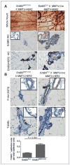
Figure 1
Figure 1
ErbB2-driven hyperplasia of the mammary epithelium requires ErbB3. A, histologic analysis by whole-mount hematoxylin staining of mammary glands harvested from 12-week-old virgin female mice revealed that diffuse epithelial hyperplasias induced upon overexpression of ErbB2 are decreased upon loss of ErbB3 (as shown by IHC) despite continued expression of ErbB2 (as shown by IHC). B, decreased phosphorylation of Akt (as shown by IHC) and increased cell death (as shown by TUNEL analysis) were evident in ErbB3-deficient mammary epithelia. Arrows indicate TUNEL+ cells. Values represent the average TUNEL+ epithelial cells per total epithelial cells, N = 4 mammary glands per genotype, 5 random fields per sample. P value calculated using Student t test.
Figure 2
Figure 2
ErbB2-expressing cells escaping Cre-mediated ErbB3 ablation give rise to ErbB3-expressing tumors in ErbB3MMTV-KO mice. A, whole-mount hematoxylin–stained mammary glands from 27-week-old virgin female mice revealed focal neoplasias in hypoplastic ErbB3MMTV-KO mammary glands. H&E-stained tumors revealed similar histologic characteristics of MMTV-HER2 tumors from ErbB3MMTV-KO and ErbB3FL/+ mice. B and C, Kaplan–Meier analysis of MMTV-HER2 (B) and MMTV-Neu (C) tumor latency in ErbB3MMTV-KO and ErbB3FL/+ mice. T50 = average tumor latency. P value calculated using log-rank test. D, IHC analysis of primary tumor sections and lung metastases revealed ErbB3 expression in tumors, but not in normal adjacent mammary epithelium (inset), of ErbB3MMTV-KO mice. E, β-galactosidase activity assessed in ErbB3MMTV-KO X MMTV-HER2 X Rosa26 mice revealed a large number of cells negative for Cre activity. F, tumor distribution across the 10 mammary glands was altered in ErbB3MMTV-KO mice as compared with ErbB3FL/+ mice, suggesting nonoverlapping expression of MMTV-Cre and MMTV-HER2 transgenes, despite the similarities in their promoter sequences.
Figure 3
Figure 3
Decreased cell growth, cell survival, hyperplasia, and tumor formation in MMTV-NIC mice, expressing ErbB2 and Cre from the same polycistronic transcript. A, histologic analysis of mammary glands harvested from 12-week-old virgin female mice by whole-mount hematoxylin staining, IHC detection of Ki67, and TUNEL analysis revealed that loss of ErbB3 in ErbB3FL/FL X NIC mice decreased diffuse hyperplasia and focal neoplasia, epithelial proliferation, and cell survival. RNA harvested from ErbB3+/+NIC (n = 8), ErbB3+/FLNIC (n = 7), and ErbB3FL/FLNIC (n = 7) was used to assess ErbB3 mRNA expression levels. Values are expressed as mean fold differences (±SE) in comparison with ErbB3+/+NIC, and normalized to the Rplp0 housekeeping gene. B, DCIS too small for detection by manual palpation became evident in 2/15 ErbB3FL/FL X NIC mice by 6 months of age. Analysis of these 2 DCIS revealed decreased cell proliferation (as seen by Ki67 IHC) and increased cell death (as seen by TUNEL analysis). Comparable DCIS lesions were evident in ErbB3FL/+ X NIC mice, as were frank adenocarcinomas, but DCIS lesions found in control samples displayed profoundly elevated cell proliferation and very low levels of cell death. Higher power magnifications of boxed areas are shown at the right. Bottom, Western analysis of whole mammary lysates using the antibodies indicated at the right of each panel. Genotype for ErbB3 is indicated as +/+ (ErbB3+/+ X NIC), FL/+ (ErbB3FL/+ X NIC) and F/FL (ErbB3FL/FL X NIC). C, Kaplan–Meier analysis of tumor latency in MMTV-NIC mice. Average tumor latency could not be determined (n.d.) in ErbB3FL/FL mice, as the majority of these mice did not develop tumors within the course of this study. Loss of a single ErbB3 allele (ErbB3FL/+ X NIC) did not affect tumor latency as compared with ErbB3+/+. D, average tumor burden per mouse, as determined by the average number of tumors per mouse (top) or total tumor volume (bottom) at 180 days of age, was measured. P value calculated using Student t test. E, lung metastases were identified upon histologic examination. Average number of lung metastases per mouse is shown, N = 8 (ErbB3+/+), 7 (ErbB3FL/+), and 15 (ErbB3FL/FL). P value, Student t test. Representative lung sections are shown.
Figure 4
Figure 4
Inducible Cre-mediated ErbB3 ablation decreases growth of ErbB2-overexpressing mammary tumors. A, treatment of 12-week-old virgin female ErbB3DOX-KO X MMTV-Neu mice with doxycycline for 14 days decreased mammary epithelial ErbB3 expression and ErbB2-induced lobuloalveolar density. B, ErbB3DOX-KO X MMTV-Neu female mice remained without doxycycline until tumors surpassed 200 mm3, at which point mice were treated with doxycycline (2 mg/mL in drinking water). Tumor volume was measured twice weekly. Values shown represent the average ± SD. P value calculated using Student t test, N = 8. C, histologic analysis of tumors treated 28 days with doxycycline show decreased tumor cellularity and increased acellular debris. D, ErbB3, P-Akt, Ki67, and cell survival were decreased in doxycycline-treated samples. TUNEL analysis revealed increased cell death in doxycycline-treated tumors. E and F, values shown indicate the average number of Ki67+ cells (E) or TUNEL+ cells (F) per total tumor cells. N = 4 per group, 5 fields per sample. P values calculated using Student t test. DOX, doxycycline.
Figure 5
Figure 5
Inhibition of ErbB3 expression or ErbB3-to-PI3K signaling improves response of ErbB2-overexpressing breast cancers to lapatinib. A, ErbB3DOX-KO mice bearing tumors more than 200 mm3 were randomized into groups treated ±doxycycline (2 mg/mL in drinking water) and ±lapatinib (100 mg/kg once daily). Tumor volume was measured twice weekly. Left, average tumor volume ± SE. Right, fold-increase in tumor volume calculated individually for each tumor, then used to calculate average fold increase in tumor volume for each group. P values, Student t test. N = 8. B, primary NiB3 cells were infected with Ad.Cre or Ad.LacZ. Cells (1 × 10⁴) were cultured in 10% serum ± lapatinib (1 μg/mL) or dimethyl sulfoxide (DMSO) for 96 hours. Viable cells were counted (Bio-Rad TC-10). Values represent average cell number ± SD. *, P < 0.01. C and D, NiB3 cells were infected with pBMN retrovirus expressing the empty vector, ErbB3WT, or ErbB36F. Cells were infected with Ad.Cre or Ad.LacZ. Ten days after infection, cell lysates were used for Western blot analysis (C) or ErbB3 immunoprecipitation followed by Western blot analysis (D). E, 24 hours after infection with Ad.Cre or Ad.LacZ, NiB3 cells (1 × 10⁴) were cultured in 10% serum for 96 hours. Viable cells were counted. Values shown represent the average ± SD, N = 3 per group analyzed in duplicate. *, P < 0.01, Student t test. F, 24 hours after infection with Ad.Cre, NiB3 cells (1 × 10⁴) were cultured with lapatinib (1 μmol/L), BKM120 (2 μmol/L), or DMSO for 21 days, stained with crystal violet and imaged. G, NiB3 tumors cells stably expressing ErbB3WT or ErbB6F were infected with Ad.Cre and cultured for 7 days. Cells (106) were injected into the left (NiB3-ErbB36F) and right (NiB3-ErbB3WT) inguinal mammary fat pads. Tumor bearing mice were treated daily 5 days with lapatinib or BKM120. TUNEL analysis of tumors collected 1 hour after final treatment was quantitated as average number of apoptotic bodies per ×400 field (N = 9–15 per group). P values were calculated using Student t test. H, representative photomicrograph of TUNEL-stained NiB3 tumors. DOX, doxycycline.

References

    1. Hynes NE, Stern DF. The biology of erbB-2/neu/HER-2 and its role in cancer. Biochim Biophys Acta. 1994;1198:165–84. - PubMed
    1. Holbro T, Civenni G, Hynes NE. The ErbB receptors and their role in cancer progression. Exp Cell Res. 2003;284:99–110. - PubMed
    1. Stern DF. ERBB3/HER3 and ERBB2/HER2 duet in mammary development and breast cancer. J Mammary Gland Biol Neoplasia. 2008;13:215–23. - PMC - PubMed
    1. Hellyer NJ, Cheng K, Koland JG. ErbB3 (HER3) interaction with the p85 regulatory subunit of phosphoinositide 3-kinase. Biochem J. 1998;333:757–63. - PMC - PubMed
    1. Holbro T, Beerli RR, Maurer F, Koziczak M, Barbas CF, 3rd, Hynes NE. The ErbB2/ErbB3 heterodimer functions as an oncogenic unit: ErbB2 requires ErbB3 to drive breast tumor cell proliferation. Proc Natl Acad Sci U S A. 2003;100:8933–8. - PMC - PubMed

Publication types