. 2012 May 18;287(21):17318-17330.
doi: 10.1074/jbc.M112.340885. Epub 2012 Mar 29.

P2X7 receptor-mediated scavenger activity of mononuclear phagocytes toward non-opsonized particles and apoptotic cells is inhibited by serum glycoproteins but remains active in cerebrospinal fluid

Affiliations

P2X7 receptor-mediated scavenger activity of mononuclear phagocytes toward non-opsonized particles and apoptotic cells is inhibited by serum glycoproteins but remains active in cerebrospinal fluid

Ben J Gu et al. J Biol Chem. .

Abstract

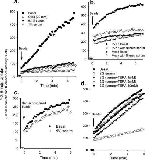
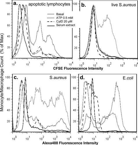
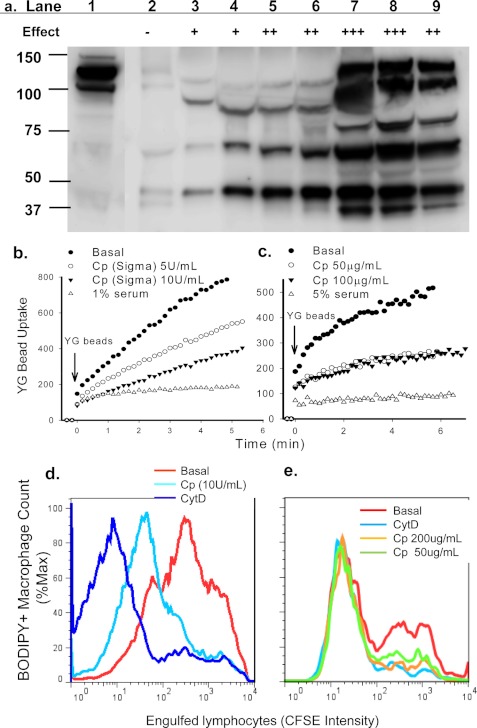
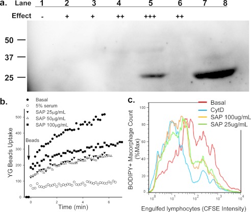
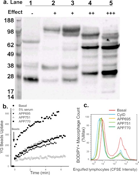
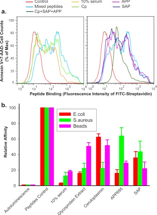
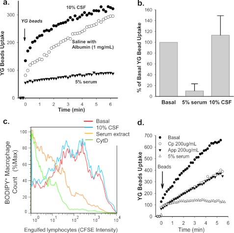
Rapid phagocytosis of non-opsonized particles including apoptotic cells is an important process that involves direct recognition of the target by multiple scavenger receptors including P2X7 on the phagocyte surface. Using a real-time phagocytosis assay, we studied the effect of serum proteins on this phagocytic process. Inclusion of 1-5% serum completely abolished phagocytosis of non-opsonized YG beads by human monocytes. Inhibition was reversed by pretreatment of serum with 1-10 mM tetraethylenepentamine, a copper/zinc chelator. Inhibitory proteins from the serum were determined as negatively charged glycoproteins (pI < 6) with molecular masses between 100 and 300 kDa. A glycoprotein-rich inhibitory fraction of serum not only abolished YG bead uptake but also inhibited phagocytosis of apoptotic lymphocytes or neuronal cells by human monocyte-derived macrophages. Three copper- and/or zinc-containing serum glycoproteins, ceruloplasmin, serum amyloid P-component, and amyloid precursor protein, were identified, and the purified proteins were shown to inhibit the phagocytosis of beads by monocytes as well as phagocytosis of apoptotic neuronal cells by macrophages. Human adult cerebrospinal fluid, which contains very little glycoprotein, had no inhibitory effect on phagocytosis of either beads or apoptotic cells. These data suggest for the first time that metal-interacting glycoproteins present within serum are able to inhibit the scavenger activity of mononuclear phagocytes toward insoluble debris and apoptotic cells.


Figures

FIGURE 1.
FIGURE 1.
Serum inhibits phagocytosis of non-opsonized beads by monocytes. Human monocytes (2 × 10⁶/ml) were labeled with APC-conjugated CD14 and incubated with 20 μm CytD for 20 min or with serum for 1 min before the addition of 1-μm YG beads. The fluorescence intensity of CD14+ monocytes was analyzed by time-resolved flow cytometry. a, a typical YG beads uptake curve shows complete inhibition of bead phagocytosis by freshly prepared human monocytes by 1% serum. The curve is representative of >10 human serum samples. b, shown is phagocytosis of YG beads by HEK-293 cells transfected with DsRed-tagged P2X7 constructs or DsRed-monomer-N1 vector (Mock) in the presence or absence of 2% serum pre-filtered through a 300-kDa ultrafiltration tube. Cells were gated on DsRed for analysis. c, uptake of YG beads opsonized by preincubation with 5% serum for 3 days at 4 °C is shown. 5% serum failed to inhibit uptake of serum-opsonized beads. d, serum pretreated with 1–10 mm TEPA showed little or no inhibition of YG bead uptake.
FIGURE 2.
FIGURE 2.
The anti-phagocytic effect of extracted serum glycoprotein-rich fraction. Human monocyte-derived macrophages (a) or fresh monocytes (b, c, and d) were pretreated with 0.5 mm ATP for 15 min, 20 μm CytD for 30 min, or ∼250 μg/ml glycoprotein-rich serum fraction for 1 min (a concentration at which the phagocytosis of non-opsonized YG beads by monocytes was completely inhibited). a, phagocytosis of CFSE-labeled apoptotic lymphocytes by autologous monocyte-derived macrophages is shown. b, phagocytosis of CFSE-labeled live S. aureus by human monocytes is shown. c and d, phagocytosis of heat-killed Alexa 488-conjugated S. aureus (c) or E. coli. (d) by human monocytes is shown.
FIGURE 3.
FIGURE 3.
CP inhibits phagocytosis of beads and apoptotic cells. a, immunoreactive CP protein in column fractions was detected by Western blotting. Lane 1, starting material; lanes 29, fractions (30 μl each) eluted from Superdex 200 gel filtration. The relative inhibitory effect of each fraction on YG bead uptake by monocytes is indicated (−, no inhibition; + to +++, increasing strength of inhibition). b and c, human PBMC labeled with APC-conjugated anti-CD14 mAb were resuspended in sodium medium with 0.1 mm Ca2+. CP or serum was added 1 min before the addition of YG beads. The fluorescence intensity of CD14+ monocytes containing engulfed beads were analyzed by time-resolved flow cytometry. d and e, flow cytometry histograms show phagocytosis of CFSE-labeled apoptotic lymphocytes by autologous monocyte-derived macrophages labeled with CMTMR or BODIPY. Mixed cells were incubated with CP or CytD (20 μm) for 3 h before cells were collected and fixed for flow cytometry assay. Inhibitors used were commercially obtained CP (∼50 unit/mg) (b and d), CP purified from human serum (c and e), or human serum (b and c).
FIGURE 4.
FIGURE 4.
SAP inhibits phagocytosis of beads and apoptotic cells. a, SAP protein in column fractions was detected by Western blotting. Lane 1–6, fractions were eluted from Superdex 200 gel filtration. The relative inhibitory effect of each fraction on YG bead uptake by monocytes is indicated; lanes 7 and 8 are 5 and 10 μl of purified SAP (1.0 mg/ml), respectively. b, human PBMC labeled with APC-conjugated anti-CD14 mAb were resuspended in sodium medium with 0.1 mm Ca2+. Purified SAP was added 1 min before the addition of YG beads. The uptake of YG beads by CD14+ monocytes was analyzed by time-resolved flow cytometry. c, flow cytometry histograms show phagocytosis of CFSE-labeled apoptotic lymphocytes by autologous monocyte-derived macrophages labeled with CMTMR or BODIPY. Mixed cells were incubated with SAP or CytD (20 μm) for 3 h before cells were collected and fixed for flow cytometry assay.
FIGURE 5.
FIGURE 5.
APP inhibits phagocytosis of beads and apoptotic cells. a, shown is APP protein expression detected by Western blotting with anti-APP mAb (clone 22C11). Lanes 15, fractions eluted from Superdex 200 gel filtration. The relative inhibitory effect of each fraction on YG bead uptake by monocytes is indicated. b, human PBMC labeled with APC conjugated anti-CD14 mAb were resuspended in sodium medium with 0.1 mm Ca2+. Recombinant APP695α, APP751α, or APP770α (100 μg/ml each) was added 1 min before the addition of YG beads. The uptake of YG beads by CD14+ monocytes were analyzed by time-resolved flow cytometry. c, flow cytometry histograms show phagocytosis of CFSE-labeled apoptotic lymphocytes by autologous monocyte-derived macrophages labeled with CMTMR or BODIPY. Mixed cells were incubated with isoforms of APP (200 μg/ml) or CytD (20 μm) for 3 h before cells were collected and fixed for flow cytometry assay.
FIGURE 6.
FIGURE 6.
Serum glycoproteins inhibit peptide binding to phagocytic target particles. Short biotin-labeled peptides, identical to the P2X7 extracellular domain sequence, show binding to the surface of apoptotic lymphocytes (a) or live E. coli, S. aureus, and 3-μm latex beads (b). Particles were incubated with mixed peptides (5 μg/ml each) in the presence or absence of 10% human serum, 0.5 mg/ml glycoprotein extract, 200 μg/ml purified CP, 200 μg/ml APP695, 100 μg/ml SAP for 15 min, or all three glycoproteins together. a, shown is a flow cytometry histogram of peptides binding. Apoptotic cells were incubated with FITC-conjugated streptavidin, APC-conjugated annexin V, and 7-AAD for 30 min. Cells were then analyzed by flow cytometry on gated Annexin V+7-AAD−/+ population. b, shown is peptide binding as measured by chemiluminescence assay. Beads and bacteria were incubation with 100 μl of HRP-labeled streptavidin (1:2000) for 15 min. Data were normalized to binding of peptide control in the absence of serum or glycoproteins and are presented as the mean ± S.D. (n = 3).
FIGURE 7.
FIGURE 7.
The effect of CSF on phagocytosis of YG beads and apoptotic neuronal cells by human monocyte/macrophages. a, a typical YG beads uptake curve shows phagocytosis of beads by human monocytes is inhibited by 5% serum but not by 10% CSF. b, composite results for YG bead uptake at 5 min from 15 CSF samples and >40 human serum samples (*, p < 0.001). c, shown is phagocytosis of apoptotic SH-SY5Y neuroblastoma cells by human monocyte-derived macrophages. SH-SY5Y cells were induced to apoptosis by 0.2 μm staurosporine for 30 min and labeled with 10 μm CFSE. Before the addition of SH-SY5Y cells, BODIPY 630/650-SE (5 μm)-labeled human macrophages were incubated with 10% CSF or 10% serum glycoprotein-rich fraction or 20 μm CytD. Mixed cells were incubated for 3 h before collection and fixation. Results are representative of eight experiments with different CSF samples. d, phagocytosis of YG beads by human monocytes is shown. Cells were resuspended in sodium medium containing 50% mixed human CSF with or without 200 μg/ml CP, 200 μg/ml APP695α, or 5% serum before the addition of YG beads.

References

    1. Cline M. J., Lehrer R. I. (1968) Phagocytosis by human monocytes. Blood 32, 423–435 - PubMed
    1. Ravichandran K. S. (2003) “Recruitment signals” from apoptotic cells. Invitation to a quiet meal. Cell 113, 817–820 - PubMed
    1. Ravichandran K. S., Lorenz U. (2007) Engulfment of apoptotic cells. Signals for a good meal. Nat. Rev. Immunol. 7, 964–974 - PubMed
    1. Wong K., Valdez P. A., Tan C., Yeh S., Hongo J. A., Ouyang W. (2010) Phosphatidylserine receptor Tim-4 is essential for the maintenance of the homeostatic state of resident peritoneal macrophages. Proc. Natl. Acad. Sci. U.S.A. 107, 8712–8717 - PMC - PubMed
    1. Park D., Tosello-Trampont A. C., Elliott M. R., Lu M., Haney L. B., Ma Z., Klibanov A. L., Mandell J. W., Ravichandran K. S. (2007) BAI-1 is an engulfment receptor for apoptotic cells upstream of the ELMO/Dock180/Rac module. Nature 450, 430–434 - PubMed

Publication types