

Role of microparticles in dengue virus infection and its impact on medical intervention strategies
- PMID: 22461739
- PMCID: PMC3313537
Role of microparticles in dengue virus infection and its impact on medical intervention strategies
Abstract
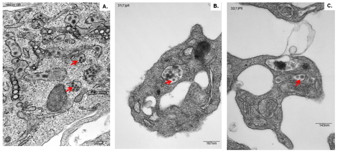
Dengue virus (DV) is one of the most important vector-borne diseases in the world. It causes a disease that manifests as a spectrum of clinical symptoms, including dengue hemorrhagic fever. DV is proficient at diverting the immune system to facilitate transmission through its vector host, Aedes spp. mosquito. Similar to other vector-borne parasites, dengue may also require a second structural form, a virus of alternative morphology (VAM), to complete its life cycle. DV can replicate to high copy numbers in patient plasma, but no classical viral particles can be detected by ultra-structural microscopy analysis. A VAM appearing as a microparticle has been recapitulated with in vitro cell lines Meg01 and K562, close relatives to the cells harboring dengue virus in vivo. VAMs are likely to contribute to the high viremia levels observed in dengue patients. This review discusses the possible existence of a VAM in the DV life cycle.
Keywords: Aedes mosquitoes; Dengue virus; alternative virion; microparticles; vaccine; vector-borne transmission.
Figures

References
-
- NIAID. Dengue Fever [Internet] 2005. Apr, Available from: http://www.niaid.nih.gov/factsheets/dengue.htm .
-
- WHO. Dengue guidelines for diagnosis, treatment, prevention and control. World Health Organization [Internet] 2009. Available from: http://whqlibdoc.who.int/publications/2009/9789241547871_eng.pdf . - PubMed
-
- Kurane I, Kontny U, Janus J, Ennis FA. Dengue-2 virus infection of human mononuclear cell lines and establishment of persistent infections. Arch Virol. 1990;110(1-2):91–101. - PubMed
-
- Cabello-Gutierrez C, Manjarrez-Zavala ME, Huerta-Zepeda A, Cime-Castillo J, Monroy-Martinez V, Correa BB. et al. Modification of the cytoprotective protein C pathway during Dengue virus infection of human endothelial vascular cells. Thromb Haemost. 2009;101(5):916–928. - PubMed
Publication types
MeSH terms
Substances
LinkOut - more resources
Full Text Sources
Medical
Miscellaneous