. 2012;7(3):e34360.
doi: 10.1371/journal.pone.0034360. Epub 2012 Mar 26.

TNF-α is involved in the abnormal thymocyte migration during experimental Trypanosoma cruzi infection and favors the export of immature cells

Affiliations

TNF-α is involved in the abnormal thymocyte migration during experimental Trypanosoma cruzi infection and favors the export of immature cells

Ana Rosa Pérez et al. PLoS One. 2012.

Abstract

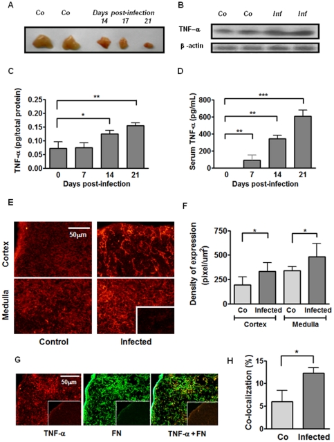
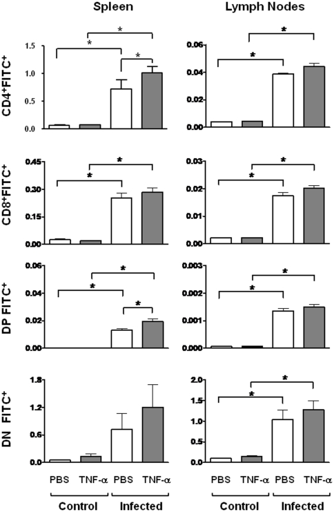
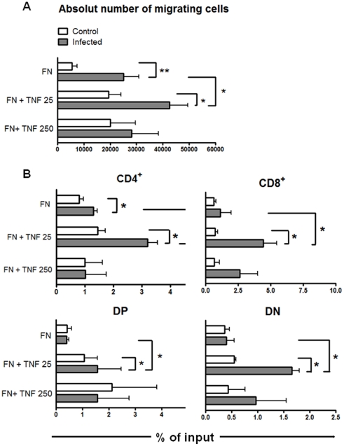
Previous studies revealed a significant production of inflammatory cytokines together with severe thymic atrophy and thymocyte migratory disturbances during experimental Chagas disease. Migratory activity of thymocytes and mature T cells seem to be finely tuned by cytokines, chemokines and extracellular matrix (ECM) components. Systemic TNF-α is enhanced during infection and appears to be crucial in the response against the parasite. However, it also seems to be involved in disease pathology, since it is implicated in the arrival of T cells to effector sites, including the myocardium. Herein, we analyzed the role of TNF-α in the migratory activity of thymocytes in Trypanosoma cruzi (T. cruzi) acutely-infected mice. We found increased expression and deposition of TNF-α in the thymus of infected animals compared to controls, accompanied by increased co-localization of fibronectin, a cell migration-related ECM molecule, whose contents in the thymus of infected mice is also augmented. In-vivo studies showed an enhanced export of thymocytes in T. cruzi-infected mice, as ascertained by intrathymic injection of FITC alone or in combination with TNF-α. The increase of immature CD4(+)CD8(+) T cells in secondary lymphoid organs was even more clear-cut when TNF-α was co-injected with FITC. Ex-vivo transmigration assays also revealed higher number of migrating cells when TNF-α was added onto fibronectin lattices, with higher input of all thymocyte subsets, including immature CD4(+)CD8(+). Infected animals also exhibit enhanced levels of expression of both mRNA TNF-α receptors in the CD4(+)CD8(+) subpopulation. Our findings suggest that in T. cruzi acute infection, when TNF-α is complexed with fibronectin, it favours the altered migration of thymocytes, promoting the release of mature and immature T cells to different compartments of the immune system. Conceptually, this work reinforces the notion that thymocyte migration is a multivectorial biological event in health and disease, and that TNF-α is a further player in the process.


Conflict of interest statement

Competing Interests: The authors have declared that no competing interests exist.

Figures

Figure 1
Figure 1. Enhanced circulating and intrathymic contents of TNF-α parallel the thymic atrophy in T. cruzi acutely-infected mice.
A) Representative picture showing two thymuses from healthy control mice (Co) and the progressive thymic atrophy after 14, 17 and 21 days of acute infection; B) TNF-α protein detection in thymus homogenates by western blot. Representative photomicrographs showing TNF-α and β-actin expression in two control mice (Co) and in two infected mice after 21 days post-infection (Inf); C) TNF-α concentrations in thymic homogenates detected by ELISA at different days after infection and normalized to total protein contents. Bars represent the mean ± SEM of three pools/day); D) Systemic concentration of TNF-α along infection evaluated by ELISA. (n = 5–8 mice/day); E) Confocal microscopy showing an enhancement in TNF-α contents both in cortex and medulla of the thymic lobules, after 14 days of infection. Original magnification 400×. Small box is a representative control staining in which an unrelated primary antibody was applied; F) Graphs correspond to relative quantification analysis of TNF-α deposition in both cortex and medulla from 3–5 microscopic fields of thymuses from control (n = 5) or 14 days-infected animals (n = 5); G) Representative immunofluorescence staining showing thymuses of infected mice: TNF-α expression (in red, left picture); fibronectin (FN) deposition (in green, middle picture); co-localization of TNF-α and FN (right picture). Small boxes correspond to negative control where immune reaction was controlled by using unrelated primary antibodies; H) Bars correspond to the percentages of TNF-α plus FN co-localization in thymuses from control mice compared with infected counterparts. Significant differences are indicated as *p<0.05, **p<0.01, ***p<0.001.
Figure 2
Figure 2. TNF-α enhances export of CD4+CD8+ thymocytes.
Control and 14 days-infected animals were intrathymically injected with FITC dissolved in PBS with or without TNF-α. Twenty-four hours later, recent thymic emigrants (RTEs) were recognized as FITC+/CD4+/CD8+ cells in secondary lymphoid organs (spleen and subcutaneous lymph nodes) by flow cytometry. Empty bars represent the absolute number of FITC+ cells (×103) from PBS injected mice and black bars represent the absolute number of FITC+ cells from TNF-α injected animals. Results show that after infection, the numbers of RTEs are clearly enhanced, independently of whether intrathymic injection of TNF-α was applied or not. When considering CD4/CD8-defined subpopulations, we found that intrathymic inoculation of TNF-α further promoted a significant enhancement in the numbers of FITC+CD4+ and FITC+CD4+CD8+ cell in the spleen, but not in lymph nodes. Data depict a representative experiment of two similar ones; each experiment being done with 5–6 animals/group. Statistically significant differences are indicated as *p<0.05.
Figure 3
Figure 3. TNF-α enhances in vitro fibronectin-driven migration of thymocytes from T. cruzi infected mice.
A) Thymocytes from control or infected animals were allowed to migrate in transwell chambers coated with fibronectin (FN) alone or FN plus TNF-α. Fibronectin-induced haptotactic response of thymocytes from infected animals was enhanced compared with controls in presence of TNF-α at the concentration of 25 pg/mL. Results derive from four experiments for TNF-25 pg/mL and three for TNF-250 pg/mL (each one obtained by pools of at least 3–4 animals by group) and correspond to specific migration after subtracting absolute cell numbers obtained in each well coated only with BSA. Statistically significant differences are indicated as p<0.05. B) Specific migration of thymocyte subpopulations was expressed as percentage of input (see Material and Methods session). We observed an enhanced migratory response of CD4+, CD8+, DP and DN cells when TNF-α was applied at 25 pg/mL, as compared with FN alone in infected animals. Results derived from four experiments, each one obtained by pools of at least 3–4 animals by group. Data correspond to specific migration after subtracting absolute cell numbers obtained in each well coated only with BSA. Statistically significant differences are indicated as *p<0.05 and **p<0.01.
Figure 4
Figure 4. Expression of TNF-R1 and TNF-R2 en CD4+, CD8+ and CD4+CD8+ (DP) thymocytes.
DP and SP T cells were purified from thymuses of control and acutely infected mice (14 days post-infection) by cell sorting using flow cytometry. To quantitatively evaluate TNFR transcripts, total mRNA samples from highly purified fresh DP, CD4+ and CD8+ T cells were processed for quantitative RT-PCR. The relative gene expression of mRNA of TNF-R1 and TNF-R2 were enhanced in DP thymocytes from infected animals compared with controls. Results are presented as relative gene expression, where mRNA levels were calculated using the equation 2−ΔCt (difference in Ct between β-actin and the target gene). Data depict a representative experiment. Statistically significant differences are indicated as *p<0.05.

Similar articles

Cited by

References

    1. Leite-de-Moraes MC, Hontebeyrie-Joskowicz M, Leboulenger F, Savino W, Dardenne M, et al. Studies on the thymus in Chagas' disease. II. Thymocyte subset fluctuations in Trypanosoma cruzi-infected mice: relationship to stress. Scand J Immunol. 1991;33:267–275. - PubMed
    1. Roggero E, Pérez A, Tamae-Kakazu M, Piazzon I, Nepomnaschy I, et al. Differential susceptibility to acute Trypanosoma cruzi infection in BALB/c and C57BL/6 mice is not associated with a distinct parasite load but cytokine abnormalities. Clin Exp Immunol. 2002;128:421–8. - PMC - PubMed
    1. Pérez AR, Roggero E, Nicora A, Palazzi J, Besedovsky HO, et al. Thymus atrophy during Trypanosoma cruzi infection is caused by an immuno-endocrine imbalance. Brain Behav Immun. 2007;21:890–900. - PubMed
    1. Cotta-de-Almeida V, Bonomo A, Mendes-da-Cruz DA, Riederer I, De Meis J, et al. Trypanosoma cruzi infection modulates intrathymic contents of extracellular matrix ligands and receptors and alters thymocyte migration. Eur J Immunol. 2003;33:2439–2448. - PubMed
    1. Mendes-da-Cruz DA, Silva JS, Cotta-de-Almeida V, Savino W. Altered thymocyte migration during experimental acute Trypanosoma cruzi infection: combined role of fibronectin and the chemokines CXCL12 and CCL4. Eur J Immunol. 2006;36:1486–1493. - PubMed

Publication types

MeSH terms

Substances