

Clinical, radiological, and genetic similarities between patients with Chiari Type I and Type 0 malformations
- PMID: 22462700
- PMCID: PMC3678957
- DOI: 10.3171/2011.12.PEDS11113
Clinical, radiological, and genetic similarities between patients with Chiari Type I and Type 0 malformations
Abstract
Object: Although Chiari Type I (CM-I) and Type 0 (CM-0) malformations have been previously characterized clinically and radiologically, there have been no studies focusing on the possible genetic link between these disorders. The goal of this study was to identify families in whom CM-0 and CM-I co-occurred and to further assess the similarities between these disorders.
Methods: Families were ascertained through a proband with CM-I. Detailed family histories were obtained to identify first-degree relatives diagnosed with CM-0. Several criteria were used to exclude individuals with acquired forms of CM-I and/or syringomyelia. Individuals were excluded with syndromic, traumatic, infectious, or tumor-related syringomyelia, as well as CM-I due to a supratentorial mass, hydrocephalus, history of cervical or cranial surgery unrelated to CM-I, or development of symptoms following placement of a lumbar shunt. Medical records and MR images were used to characterize CM-I and CM-0 individuals clinically and radiologically.
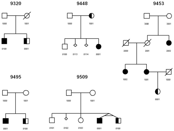
Results: Five families were identified in which the CM-I proband had a first-degree relative with CM-0. Further assessment of affected individuals showed similar clinical and radiological features between CM-0 and CM-I individuals, although CM-I patients in general had more severe symptoms and skull base abnormalities than their CM-0 relatives. Overall, both groups showed improvement in symptoms and/or syrinx size following craniocervical decompression surgery.
Conclusions: There is accumulating evidence suggesting that CM-0 and CM-I may be caused by a common underlying developmental mechanism. The data in this study are consistent with this hypothesis, showing similar clinical and radiological features between CM-0 and CM-I individuals, as well as the occurrence of both disorders within families. Familial clustering of CM-0 and CM-I suggests that these disorders may share an underlying genetic basis, although additional epigenetic and/or environmental factors are likely to play an important role in the development of CM-0 versus CM-I.
Figures
References
-
- Atkinson JL, Kokmen E, Miller GM. Evidence of posterior fossa hypoplasia in the familial variant of adult Chiari I malformation: case report. Neurosurgery. 1998;42:401–404. - PubMed
-
- Boyles AL, Enterline DS, Hammock PH, Siegel DG, Slifer SH, Mehltretter L, et al. Phenotypic definition of Chiari type I malformation coupled with high-density SNP genome screen shows significant evidence for linkage to regions on chromosomes 9 and 15. Am J Med Genet A. 2006;140:2776–2785. - PubMed
-
- Cavender RK, Schmidt JH., III Tonsillar ectopia and Chiari malformations: monozygotic triplets. Case report. J Neurosurg. 1995;82:497–500. - PubMed
-
- Chern JJ, Gordon AJ, Mortazavi MM, Tubbs RS, Oakes WJ. Pediatric Chiari malformation Type 0: a 12-year institutional experience. Clinical article. J Neurosurg Pediatr. 2011;8:1–5. - PubMed
Publication types
MeSH terms
Grants and funding
LinkOut - more resources
Full Text Sources
Medical
