. 2012 Apr 1:12:30.
doi: 10.1186/1471-230X-12-30.

Glucagon-like peptides 1 and 2 and vasoactive intestinal peptide are neuroprotective on cultured and mast cell co-cultured rat myenteric neurons

Affiliations

Glucagon-like peptides 1 and 2 and vasoactive intestinal peptide are neuroprotective on cultured and mast cell co-cultured rat myenteric neurons

Ulrikke Voss et al. BMC Gastroenterol. .

Abstract

Background: Neuropathy is believed to be a common feature of functional and inflammatory intestinal diseases. Vasoactive intestinal peptide (VIP) is an acknowledged neuroprotective agent in peripheral, including enteric, and central neurons. The proglucagon-like hormones glucagon-like peptide 1 and 2 (GLP1 and GLP2) belong to the secretin/glucagon/VIP superfamily of peptides and GLP1 and GLP2 receptors are expressed in enteric neurons. Possible neuroprotective effects of these peptides were investigated in the present study.

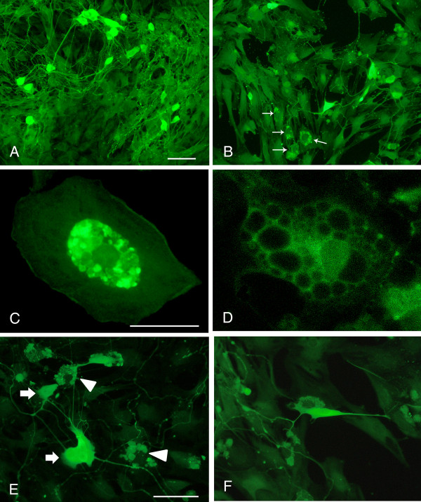
Methods: GLP1, GLP2 and VIP were added to cultured myenteric neurons from rat small intestine or to co-cultures of myenteric neurons and rat peritoneal mast cells. Receptor selectivity was tested by the simultaneous presence of a GLP1 receptor antagonist (exendin (9-39) amide) or a VIP receptor antagonist (hybrid of neurotensin 6-11 and VIP 7-28). Neuronal survival was examined using immunocytochemistry and cell counting.

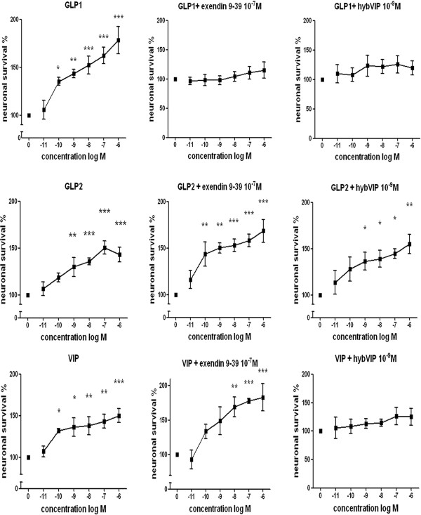
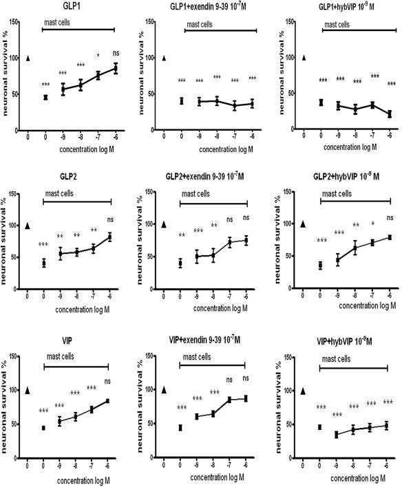
Results: GLP1, GLP2 and VIP significantly and concentration-dependently enhanced neuronal survival. In addition the peptides efficiently counteracted mast cell-induced neuronal cell death in a concentration-dependent manner. Exendin(9-39)amide reversed GLP1-induced neuroprotection while GLP2- and VIP-induced enhanced neuronal survival were unaffected. The VIP receptor antagonist reversed GLP1- and VIP-induced neuroprotection while the GLP2-induced effect on neuronal survival was unaffected.

Conclusions: By activating separate receptors VIP, GLP1 and GLP2 elicit neuroprotective effects on rat myenteric neurons cultured with or without mast cells. This implies a powerful therapeutic potential of these peptides in enteric neuropathies with a broad spectrum of applications from autoimmunity to functional disorders.


Figures

Figure 1
Figure 1
Immunocytochemical staining of myenteric neurons cultured in absence (A) or presence (B-F) of peritoneal mast cells. A. Neurons grown for 6 days and stained with PGP9.5. The cultured neurons survive well; they group into ganglion-like structures and grow a prominent arborizing network of nerve terminals. B. Neurons co-cultured with mast cells (arrows) stained with antibodies against PGP9.5 and histamine, respectively. In the neuronal cultures to which mast cells have been added the number of neurons markedly decreases and the terminal network is disintegrated. C and D. show mast cells grown in co-culture and stained with histamine antibodies. The mast cells are well settled within the cultures and they contain a high number of cytoplasmic granules. C. illustrates a cultured mast cell with numerous granules in the central, perinuclear, portion of the cell. D. shows a mast cell exhibiting piece meal degranulation. E and F. illustrate the morphological relationship of cultured neurons and mast cells. E. shows mast cells (arrowheads) in close contact with nerve terminals from nearby myenteric neurons (arrows). F. a mast cell and a nerve cell body in close proximity. In order to perceive the morphological arrangement of mast cells and neurons both PGP9.5- and histamine- immunoreactive cells are visualised by FITC conjugated antibodies. Neurons and mast cells are distinguished by their morphological characteristics. Bar in A 100 μm represents also B, bar in C 40 μm represents also D, bar in E 50 μm represents also F.
Figure 2
Figure 2
Survival of myenteric neurons pre-cultured for 4 days followed by 2 days of culture in various concentrations (10-11- 10-6 M) of GLP1, GLP2 or VIP in the absence or presence of GLP1 receptor antagonist (exendin(9-39)amide, 10-7 M) or VIP receptor antagonist (hybVIP, 10-9 M). Presence of GLP1, GLP2 and VIP significantly and concentration-dependently increase neuronal survival. Simultaneous addition of exendin(9-39)amide attenuates GLP1-induced neuroprotection while that of GLP2 and VIP are unaffected. Simultaneous addition of hybVIP attenuates GLP1- and VIP-induced neuroprotection while that of GLP2 is unaffected. Means ± SEM, n = 6-21; * p < 0.1; ** p < 0.05; *** p < 0.01 as compared to controls run in parallel without addition of peptides or antagonists.
Figure 3
Figure 3
Survival of neurons pre-cultured for 4 days followed by 2 days of culture in the absence (▲) or presence of mast cells (■, indicated by horizontal bar in each graph). During the co-culture period the effects of GLP1, GLP2 and VIP (10-9- 10-6 M), in the absence or presence of GLP1 receptor antagonist (exendin(9-39)amide, 10-7 M) or VIP receptor antagonist (hybVIP, 10-7 M), on neuronal survival were determined by neuronal cell counting. With increasing concentrations all three peptides were able to reverse mast cell-induced neuronal cell death (denoted ns; no significant difference). Simultaneous presence of exendin(9-39)amide (10-7 M) counteracted the effects of GLP1 while GLP2- or VIP-induced neuroprotection was unaffected. Addition of hybVIP (10-9 M) to the co-cultures hampered GLP1- and VIP-, but not GLP2-, induced neuroprotection. Means ± SEM, n = 5-12; ns, no significant difference; * p < 0.1; ** p < 0.05; *** p < 0.01 as compared to controls run in parallel (▲) without addition of mast cells, peptides or antagonists.

References

    1. Hammar J, Howell S, Bytzer P, Horowitz M, Talley NJ. Symptom clustering in subjects with and without diabetes mellitus: A population-based study of 15 000 Australian adults. Am J Gastroenterol. 2003;98:391–398. - PubMed
    1. Chandrasekharan B, Srinivasan S. Diabetes and the enteric nervous system. Neurogastroenterol Motil. 2007;19:951–960. - PMC - PubMed
    1. Guo C, Quobatari A, Shangguan Y, Hong S, Wiley JW. Diabetic autonomic neuropathy: evidence for apoptosis in situ in the rat. Neurogastroenterol Motil. 2004;16:335–345. doi: 10.1111/j.1365-2982.2004.00524.x. - DOI - PubMed
    1. Shotton HR, Broadbent S, Lincoln J. Prevention and partial reversal of diabetes-induced changes in enteric nerves of rat ileum by combined treatment with alpha-lipoic acid and evening primrose oil. Autonom Neurosci. 2004;111:57–65. doi: 10.1016/j.autneu.2004.02.004. - DOI - PubMed
    1. Voukali E, Shotton HR, Lincoln J. Selective responses of myenteric neurons to oxidative stress and disbetic stimuli. Neurogastroenterol Motil. 2011;23:964–e411. doi: 10.1111/j.1365-2982.2011.01778.x. - DOI - PubMed

Publication types

LinkOut - more resources