. 2012 Nov 16:1485:63-76.
doi: 10.1016/j.brainres.2012.03.016. Epub 2012 Mar 13.

Amygdala hyperactivity and tonotopic shift after salicylate exposure

Affiliations

Amygdala hyperactivity and tonotopic shift after salicylate exposure

Guang-Di Chen et al. Brain Res. .

Abstract

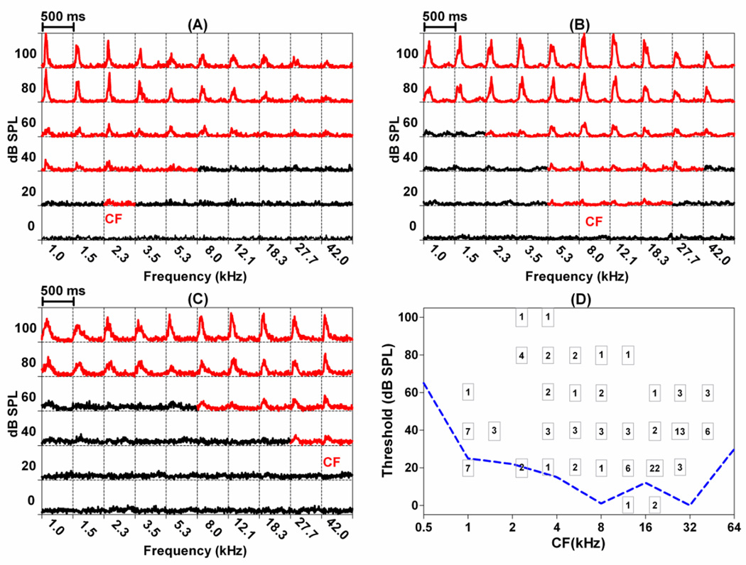
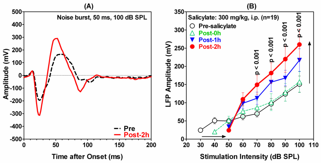
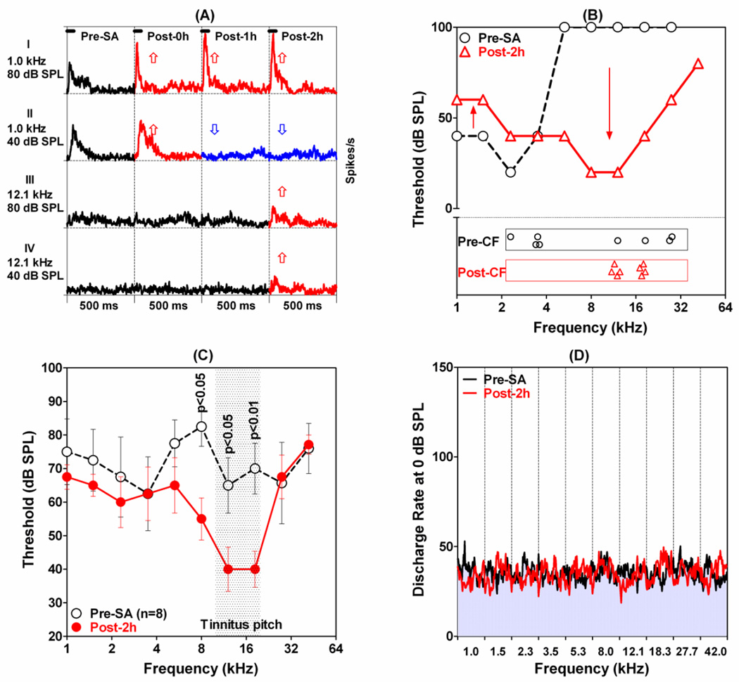
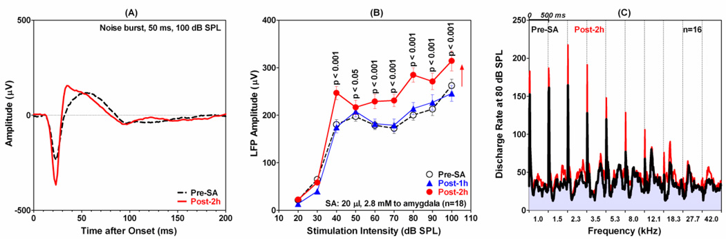
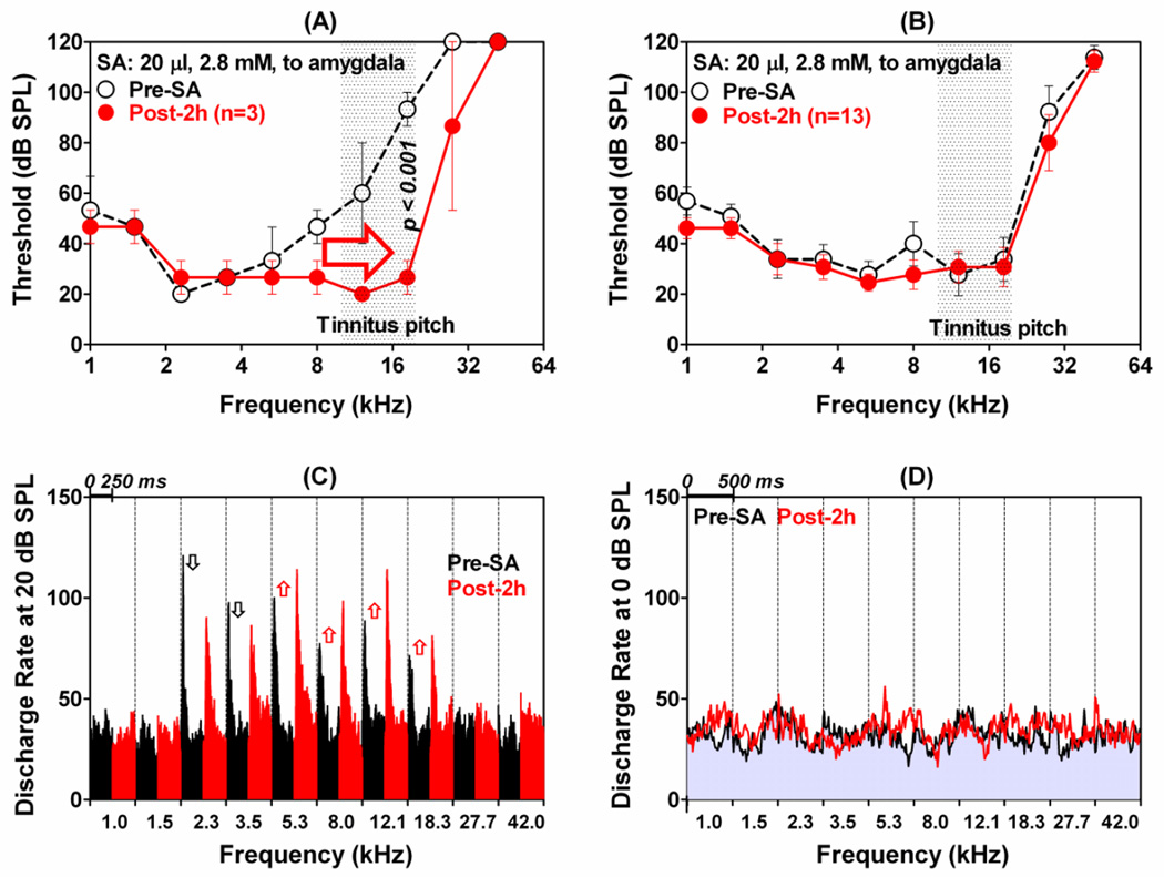
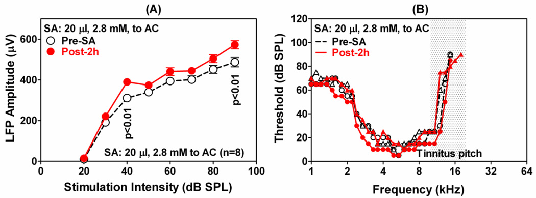
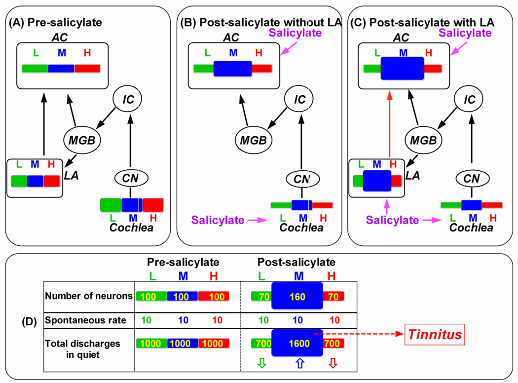
The amygdala, important in forming and storing memories of aversive events, is believed to play a major role in debilitating tinnitus and hyperacusis. To explore this hypothesis, we recorded from the lateral amygdala (LA) and auditory cortex (AC) before and after treating rats with a dose of salicylate that induces tinnitus and hyperacusis-like behavior. Salicylate unexpectedly increased the amplitude of the local field potential (LFP) in the LA making it hyperactive to sounds≥60 dB SPL. Frequency receptive fields (FRFs) of multiunit (MU) clusters in the LA were also dramatically altered by salicylate. Neuronal activity at frequencies below 10 kHz and above 20 kHz was depressed at low intensities, but was greatly enhanced for stimuli between 10 and 20 kHz (frequencies near the pitch of the salicylate-induced tinnitus in the rat). These frequency-dependent changes caused the FRF of many LA neurons to migrate towards 10-20 kHz thereby amplifying activity from this region. To determine if salicylate-induced changes restricted to the LA would remotely affect neural activity in the AC, we used a micropipette to infuse salicylate (20 μl, 2.8 mM) into the amygdala. Local delivery of salicylate to the amygdala significantly increased the amplitude of the LFP recorded in the AC and selectively enhanced the neuronal activity of AC neurons at the mid-frequencies (10-20 kHz), frequencies associated with the tinnitus pitch. Taken together, these results indicate that systemic salicylate treatment can induce hyperactivity and tonotopic shift in the amygdala and infusion of salicylate into the amygdala can profoundly enhance sound-evoked activity in AC, changes likely to increase the perception and emotional salience of tinnitus and loud sounds. This article is part of a Special Issue entitled: Tinnitus Neuroscience.


Conflict of interest statement

The authors declare no conflict of interest.

Figures

Figure 1
Figure 1
Representative frequency-receptive fields of clusters recorded in the LA: (A) low-CF, (B) mid-CF, and (C) high-CF. Each square contains a PSTH (500 ms duration, 5 ms bin width) in response to tone bursts (50 ms duration) presented at the frequency and intensity indicated on the abscissa and ordinate respectively. PSTHs in red showed an increase in firing rate during the stimulus. (D) Number inside each square shows the number of MU clusters with a CF at the indicated frequency (abscissa) and threshold (ordinate). CF: characteristic frequency; FRF: frequency receptive field; PSTH: peristimulus time histogram. Dashed line shows rat behavioral audiogram (Heffner et al., 1994).
Figure 2
Figure 2
Effect of systemic salicylate injection on LFP recorded from the amygdala in response to 50 ms noise burst. (A) A representative LFP waveform pre-salicylate (black dashed line) and 2 h following salicylate (red solid line); noise burst intensity 100 dB SPL. (B) Mean (±SEM; n=19) LFP amplitudes as a function of noise burst (50 ms) intensity before (open circle, black line) and 0 h, 1 h and 2 h after systemic administration of 300 mg/kg (i.p.) salicylate. Note significant amplitude enhancement (black vertical arrow) at intensities ≥60 dB SPL; salicylate increased thresholds approximately 20 dB (black horizontal arrow) and reduced LFP amplitudes at intensities below 60 dB SPL.
Figure 3
Figure 3
The effects of system salicylate (300 mg/kg, i.p.) on neurons in the LA. (A): Typical changes observed in PSTHs (tone bursts, 500 ms duration, 5 ms bin width) recorded from MU clusters in the LA before (left column) and 0 h, 1 h and 2 h after salicylate treatment (second, third and fourth column from left). Row I (80 dB SPL, 1 kHz): Note enhanced firing rate (red PSTHs, up arrow) after salicylate injection. Row II (1 kHz, 40 dB SPL): PSTH showed a slight enhancement (red) shortly after salicylate treatment (red PSTH, up arrow) followed by a large decrease in PSTHs amplitude (blue PSTHs, down arrow) 1 h and 2 h post-salicylate. Row III (12.1 kHz, 80 dB SPL) and Row IV (12.1 kHz, 40 dB SPL): PSTHs showed little or no response before salicylate and 0 and 1 h post-salicylate (black PSTHs); note increase in PSTH amplitude 2 h post-salicylate (red). (B) FRF of the MU cluster shown in panel A before (open circle dashed line) and 2 h post-salicylate (red open triangle, solid line). Salicylate caused an increase in low-frequency thresholds (up arrow), decrease in high-frequency thresholds (down arrow), migration of FRF toward the high frequencies and up-shift in CF. Lower portion of panel B shows CFs of 8 MU cluster in LA pre-salicylate (black open circles) and 2 h post-salicylate (red open triangles). Note up-shift of low CFs and down shift of high CF MU clusters into the 10-20 kHz range. (C) Mean (±SEM) FRFs of all 8 MU clusters (10 test frequencies, 1 to 42 kHz, 50 ms tone bursts) before (open black circles, dashed line) and 2 h post-salicylate (filled red circles, solid line). Salicylate: 300 mg/kg (i.p.). (D). Each column shows the mean PSTHs of all 8 MU clusters measured at 0 dB SPL (subthreshold) at 10 test frequencies from 1 to 42 kHz. Mean firing rate at subthreshold intensity essentially unchanged after 300 mg/kg salicylate (i.p.).
Figure 4
Figure 4
The effect of infusing 20 µl of 2.8 mM salicylate into the LA on the response properties of the AC. (A) Representative LFP waveforms to 50 ms tone burst presented at 100 dB SPL before (black dash line) and 2 h post-salicylate (red solid line). Note increase in LFP amplitude post-salicylate. (B): Mean (n=18, ±SEM) LFP amplitudes as a function of noise burst intensity (50 ms) before (open circles, dashed line) and 1 h (filled diamonds) and 2 h post-salicylate (filled circles). Note significant enhancement of LFP amplitudes from 40-100 dB SPL and lack of threshold shift. (C) Mean (n=16) PSTHs (500 ms duration, 10 ms bin width) to 50 ms tone bursts presented at 80 dB SPL at frequencies from 1 to 42 kHz. Pre-salicylate PSTHs (black line/gray body) superimposed on red PSTHS (2 h post-salicylate). Tone-evoked firing rates 2 h post-salicylate are higher than pre-salicylate firing rates, i.e., red regions above gray-black PSTHs show increases in firing rate.
Figure 5
Figure 5
The effects of infusing 20 µl of 2.8 mM of salicylate into LA on neuronal activity in AC. (A): Averaged (±SEM) FRFs of three low-CF MU clusters in the AC before (open circles, dashed line) and 2 h post-salicylate (filled circles, solid line). Note decrease in threshold and expansion along high-frequency side of the FRFs after infusing salicylate into the LA. (B): Averaged (±SEM) FRF of 13 MU clusters in the AC that showed no change in their frequency-threshold tuning after infusing salicylate into the LA. (C): Mean PSTHs (250 ms duration, 1 ms bin width) of the 13 MU clusters in panel B to tone bursts presented at 20 dB SPL. Each column shows the mean PSTH before (black, left) and 2 h post-salicylate (red, right) at frequencies from 1 to 42 kHz. Note increase in mean discharge rates from 5.3 to 18.3 kHz (up arrows) and decreased firing rates at 2.3 and 3.5 kHz (down arrows). (D) Mean PSTHs (500 ms duration, 10 ms bin width) for 13 MU clusters to tone bursts presented at 0 dB SPL (~20 below mean AC thresholds). Mean PSTHs before (gray with black line) and 2 h after (red line) salicylate infused into LA. Each column shows the mean PSTH at frequencies from 1 to 42 kHz; mean discharge rates at subthreshold intensity largely unchanged by salicylate.
Figure 6
Figure 6
Change in AC activity when salicylate was applied to the AC. (A): Mean (n=8, ±SEM) LFP amplitudes as a function of noise burst (50 ms) intensity; significant changes in amplitude indicated on the graph. (B): Representative FRFs of 2 AC neurons pre-salicylate (black) and 2 h post-salicylate (red). One MU cluster showed a slight FRF-expansion on both sides (opened and filled circles) and one which did not (open and filled triangles). Salicylate: 20 µl at 2.8 mM. Stimulation: tone bursts of 50 ms.
Figure 7
Figure 7
Highly simplified scheme showing the lateral amygdala (LA) and the auditory pathway that includes the cochlea, cochlear nucleus (CN), inferior colliculus (IC), medial geniculate body (MGB) and auditory cortex (AC). Tonotopy of the cochlea, LA and AC is indicated by bars in different colors. Extent of cochlea, LA and AC is occupied by low (L = green), middle (M = blue) and high (H = red) frequencies indicated by width of rectangle; magnitude of excitation within each frequency region indicated by height of rectangle. (A) Pre-salicylate condition showing region extent and magnitude of activity with the cochlea, LA and AC. (B) Post-salicylate scheme with the LA removed. Salicylate causes overall decrease in activity in cochlea; decreases is greatest at low and high frequencies and least at mid-frequencies (10-20 kHz). Note expansion of mid-frequency region in AC and moderate increase in excitation. (C) Post-salicylate scheme including LA. Same as in panel B except that now salicylate also increases activity in mid-frequency region of LA; hyperactivity in mid-frequency region of LA sent to corresponding region of AC further enhancing activity in the mid-frequency region of the AC. (D) Relative discharges (total discharges) in quite in the central auditory system (neuron number x spontaneous discharge rate) are equivalent across tonotopic regions under normal condition (pre-salicylate). However, an increased number of discharges in the mid-frequency region (up arrow) and a decreased number of discharges in the low- and high-frequency regions (down arrows) are expected after a high dose of salicylate due to the change of numbers of neurons which tune to the frequency. The relatively more discharges in the mid-frequency region in quiet may be accepted as a sound stimulation --- tinnitus.
Figure 8
Figure 8
(A) Photomicrograph of a coronal section through the left side of the brain showing DiI labeling (red) of the electrode track through the striatum and into the dorsal division of the lateral amygdala (LA). Neurons immunolabeled with a monoclonal antibody against NeuN and fluorescently-conjugated secondary antibody against Alexa Fluor 488 (green). (B) Higher magnification of adjacent section from the same animal as in (A) showing electrode tract in the LA. AP: anterior-posterior refer to the Bregma; ML: middle-lateral refer to the middle line.

References

    1. Amano T, Unal CT, Pare D. Synaptic correlates of fear extinction in the amygdala. Nature neuroscience. 2010;13:489–494. - PMC - PubMed
    1. Andersson G, et al. Regional cerebral blood flow during tinnitus: a PET case study with lidocaine and auditory stimulation. Acta Otolaryngol. 2000;120:967–972. - PubMed
    1. Andersson G, et al. Tinnitus distress, anxiety, depression, and hearing problems among cochlear implant patients with tinnitus. Journal of the American Academy of Audiology. 2009;20:315–319. - PubMed
    1. Arnold W, et al. Focal metabolic activation in the predominant left auditory cortex in patients suffering from tinnitus: a PET study with [18F]deoxyglucose. ORL J Otorhinolaryngol Relat Spec. 1996;58:195–199. - PubMed
    1. Bordi F, LeDoux J. Sensory tuning beyond the sensory system: an initial analysis of auditory response properties of neurons in the lateral amygdaloid nucleus and overlying areas of the striatum. J Neurosci. 1992;12:2493–2503. - PMC - PubMed

Publication types

Substances