An official website of the United States government
The .gov means it’s official.
Federal government websites often end in .gov or .mil. Before
sharing sensitive information, make sure you’re on a federal
government site.
The site is secure.
The https:// ensures that you are connecting to the
official website and that any information you provide is encrypted
and transmitted securely.
1 Division of Hematology-Oncology, Department of Medicine, Abramson Cancer Center, Abramson Family Cancer Research Institute, Perelman School of Medicine, University of Pennsylvania, Philadelphia, PA 19104, USA. dangvchi@upenn.edu
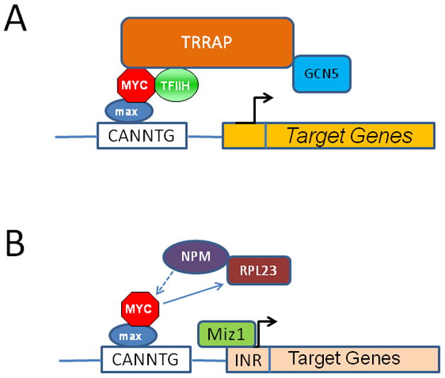
The MYC oncogene contributes to the genesis of many human cancers. Recent insights into its expression and function have led to therapeutic opportunities. MYC's activation by bromodomain proteins could be inhibited by drug-like molecules, resulting in tumor inhibition in vivo. Tumor growth can also be curbed by pharmacologically uncoupling bioenergetic pathways involving glucose or glutamine metabolism from Myc-induced cellular biomass accumulation. Other approaches to halt Myc on the path to cancer involve targeting Myc-Max dimerization or Myc-induced microRNA expression. Here the richness of our understanding of MYC is reviewed, highlighting new biological insights and opportunities for cancer therapies.
A . The MYC protooncogene is depicted downstream of receptor signal transduction pathways,…
Figure 1
A. The MYC protooncogene is depicted downstream of receptor signal transduction pathways, which elicit positive or negative regulation of the MYC gene. MYC produces the transcription factor Myc, which dimerizes with Max and bind target DNA sequences or E-boxes (with the sequence 5′-CANNTG-3′) to regulate transcription of genes involved in cell growth and proliferation. The WNT pathway is depicted with APC negatively regulating β-catenin, which upon nuclear translocation participates in the transactivation of MYC, such that loss of APC results in constitutive oncogenic MYC expression. B. When MYC is deregulated, by gene amplication, chromosomal translocation or loss of upstream regulators, such as APC, acute sustained oncogenic MYC expression results in checkpoint activation p53 or Arf. Loss of checkpoint regulation through mutations of p53 or Arf, for example, uncloaks MYC’s full tumorigenic potential.
Figure 1
A . The MYC protooncogene…
Figure 1
A . The MYC protooncogene is depicted downstream of receptor signal transduction pathways,…
Figure 1
A. The MYC protooncogene is depicted downstream of receptor signal transduction pathways, which elicit positive or negative regulation of the MYC gene. MYC produces the transcription factor Myc, which dimerizes with Max and bind target DNA sequences or E-boxes (with the sequence 5′-CANNTG-3′) to regulate transcription of genes involved in cell growth and proliferation. The WNT pathway is depicted with APC negatively regulating β-catenin, which upon nuclear translocation participates in the transactivation of MYC, such that loss of APC results in constitutive oncogenic MYC expression. B. When MYC is deregulated, by gene amplication, chromosomal translocation or loss of upstream regulators, such as APC, acute sustained oncogenic MYC expression results in checkpoint activation p53 or Arf. Loss of checkpoint regulation through mutations of p53 or Arf, for example, uncloaks MYC’s full tumorigenic potential.
Figure 2
A . The Myc-Max heterodimer…
Figure 2
A . The Myc-Max heterodimer is shown to interact with key co-factors such…
Figure 2
A. The Myc-Max heterodimer is shown to interact with key co-factors such as TFIIH that triggers transcriptional elongation or TRRAP that recruits the GCN5, which acetylates histone, permitting transcription of target genes. B. Myc-Max also mediates gene repression. Miz-1 is shown tethered to the INR element to regulate transcription of target genes, which could be silenced by Myc displacement of NPM, a Miz-1 cofactor, or by Myc induction of the ribosomal protein RPL23, which retains NPM in the nucleolus, keeping it away from Miz-1.
Figure 3
Myc regulates a network of…
Figure 3
Myc regulates a network of microRNAs through activation of the miR-17-92 cluster and…
Figure 3
Myc regulates a network of microRNAs through activation of the miR-17-92 cluster and repression of dozens of miRs including Let-7, which was recently shown to affect insulin signaling, miR-23a/b, which regulates glutaminase expression, and miR-34a, which was shown to regulate lactate dehydrogenase (LDHA) expression. The miR-17 cluster contains microRNAs that targets PTEN, thereby activating AKT, and those that targets the proapoptotic BimL or the transcription factor E2F1 expression. MicroRNAs downstream of Myc have also been implicated in epithelial-mesenchymal transition and angiogenesis.
Figure 4
Myc-Max is shown bound to…
Figure 4
Myc-Max is shown bound to E-box driven genes, which could also be regulated…
Figure 4
Myc-Max is shown bound to E-box driven genes, which could also be regulated by other E-box transcription factors, such as the carbohydrate response element binding protein (ChREBP), sterol response element binding protein (SREBP), nuclear respiratory factor 1 (NRF1), circadian transcription factor Clock (and Bmal), and hypoxia inducible factor (HIF). The non-Myc E-box transcription factors regulate genes involved in metabolism, which is maintained for cellular homeostasis when cells are not proliferating. Upon activation of MYC and elevated levels of Myc, mass action favors the binding of Myc-Max to E-box genes to regulate metabolism and genes involved in ribosomal biogenesis and cell mass accumulation. This model suggests that resting cells express metabolic genes through ‘homeostatic’ E-box transcription factors, which regulate a set of genes that overlaps with Myc target genes that are expressed when cells are stimulate to grow and proliferate.
Adams JM, Harris AW, Pinkert CA, Corcoran LM, Alexander WS, Cory S, Palmiter RD, Brinster RL. The c-myc oncogene driven by immunoglobulin enhancers induces lymphoid malignancy in transgenic mice. Nature. 1985;318:533–538.
-
PubMed
Adhikary S, Marinoni F, Hock A, Hulleman E, Popov N, Beier R, Bernard S, Quarto M, Capra M, Goettig S, et al. The ubiquitin ligase HectH9 regulates transcriptional activation by Myc and is essential for tumor cell proliferation. Cell. 2005;123:409–421.
-
PubMed
Aguda BD, Kim Y, Piper-Hunter MG, Friedman A, Marsh CB. MicroRNA regulation of a cancer network: consequences of the feedback loops involving miR-17-92, E2F, and Myc. Proc Natl Acad Sci U S A. 2008;105:19678–19683.
-
PMC
-
PubMed
Ahmadiyeh N, Pomerantz MM, Grisanzio C, Herman P, Jia L, Almendro V, He HH, Brown M, Liu XS, Davis M, et al. 8q24 prostate, breast, and colon cancer risk loci show tissue-specific long-range interaction with MYC. Proc Natl Acad Sci U S A. 2010;107:9742–9746.
-
PMC
-
PubMed