Review
. 2012 Mar 30;149(1):36-47.
doi: 10.1016/j.cell.2012.03.009.

Malignant glioma: lessons from genomics, mouse models, and stem cells

Affiliations
Review

Malignant glioma: lessons from genomics, mouse models, and stem cells

Jian Chen et al. Cell. .

Abstract

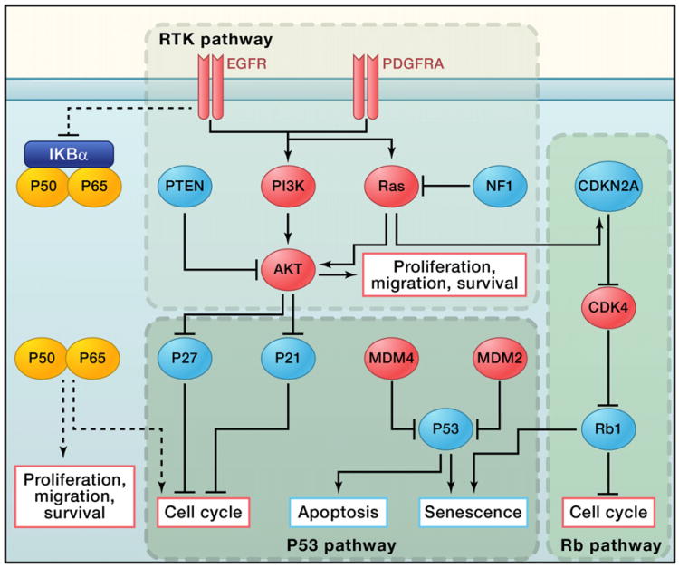
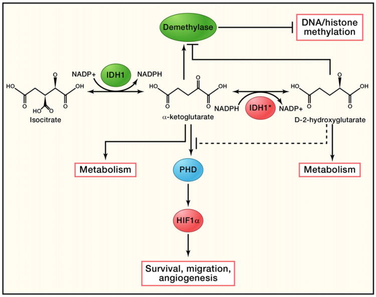
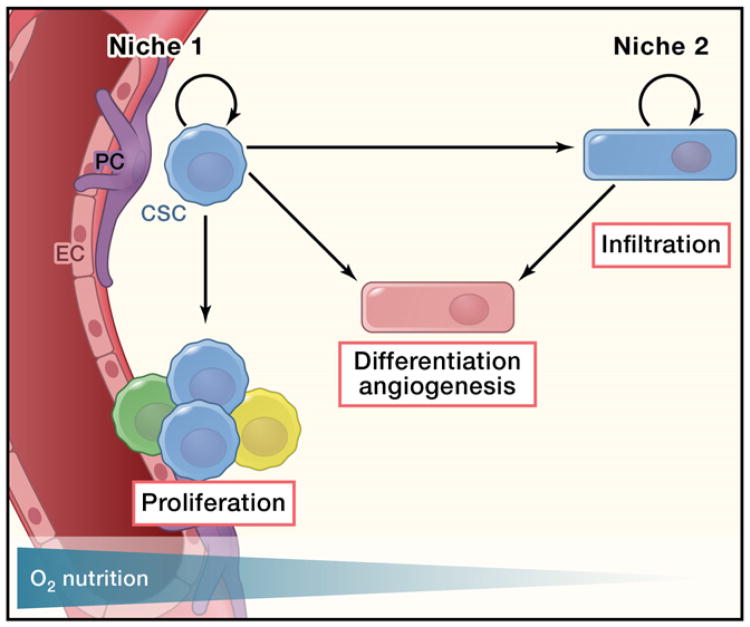
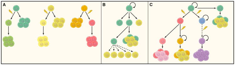
Eighty percent of malignant tumors that develop in the central nervous system are malignant gliomas, which are essentially incurable. Here, we discuss how recent sequencing studies are identifying unexpected drivers of gliomagenesis, including mutations in isocitrate dehydrogenase 1 and the NF-κB pathway, and how genome-wide analyses are reshaping the classification schemes for tumors and enhancing prognostic value of molecular markers. We discuss the controversies surrounding glioma stem cells and explore how the integration of new molecular data allows for the generation of more informative animal models to advance our knowledge of glioma's origin, progression, and treatment.


Figures

Figure 1
Figure 1. Core Signaling Pathways in Glioma Tumorigenesis
The receptor tyrosine kinase (RTK), p53, and Rb pathways are the core signaling pathways in glioma oncogenesis. Red indicates oncogenes that are either overexpressed or amplified in GBM samples, and blue indicates tumor suppressor genes that are somatically mutated or deleted (except for P27 and P21).
Figure 2
Figure 2. Function of Normal and Mutated IDH1
Wild-type IDH1 catalyzes isocitrate to form α-ketoglutarate and convert NADP+ to NADPH at the same time. A mutated form of IDH1 can convert α-ketoglutarate to D2-hydroxyglutarate in an NADPH-dependent manner. Excessive D2-hy-droxyglutarate results in cellular stress as well as metabolic changes. It could also potentially act as a competitive substrate to inhibit DNA/histone methyltransferases and prolyl hydroxylases (PHDs), resulting in DNA/histone hypomethylation or activation of HIF-1α, which can be further accelerated by the lack of α-ketoglutarate, as α-ketoglutarate is a key substrate for both PHDs and DNA/histone methyltransferases.
Figure 3
Figure 3. The Vascular Niche and Hypoxic Niche of Glioma Stem Cells
Vascular niches have been found to be important for glioma growth, probably due to the secreted factors from the endothelial cells within the niche, as well as the nutrient supply from the blood vessels. On the other hand, constantly infiltrating glioma stem cells (GSCs) probably maintain their stemness through activation of hypoxia-related pathways. At the same time, GSCs can recruit endothelial cells by secreting angiogenic factors or by directly differentiating into cells of the endothelial lineage, which in turn support glioma growth. EC and PC are endothelial cell and pericyte cell, respectively.
Figure 4
Figure 4. Cancer Maintenance Models
(A) Clonal evolution model. In this model, tumor cells are equivalent, and a majority of the tumor cells have the ability to sustain tumor growth. (B) Traditional view of the cancer stem cell model. In this model, a stable hierarchy exists in the cells of the tumor, whereby only cancer stem cells have the ability to self-renew and contribute to long-term maintenance of tumor growth. (C) Evolutional view of cancer stem cell model. This model posits that the hierarchical structure of cancer stem cells is constantly evolving due to natural selection and genomic instability. New cancer stem cell clones with different genetic alterations emerge over time. Certain genetic events will ultimately confer most tumor cells with self-renewal capacity without the reliance of niches or stemness factors (dashed arrows).
Figure 5
Figure 5. Cell of Origin of Gliomas
Mutations in adult neural stem cells are sufficient to drive malignant glioma formation in vivo. Some evidence for progenitors or mature astrocytes as the cells of origin has been demonstrated in vitro and in vivo. However, strict proof has not yet been shown due to the lack of specific in vivo lineage markers.

References

    1. Aguirre AA, Chittajallu R, Belachew S, Gallo V. NG2-expressing cells in the subventricular zone are type C-like cells and contribute to inter neuron generation in the postnatal hippocampus. J Cell Biol. 2004;165:575–589. - PMC - PubMed
    1. Alcantara Llaguno S, Chen J, Kwon CH, Jackson EL, Li Y, Burns DK, Alvarez-Buylla A, Parada LF. Malignant astrocytomas originate from neural stem/progenitor cells in a somatic tumor suppressor mouse model. Cancer Cell. 2009;15:45–56. - PMC - PubMed
    1. Anido J, Sáez-Borderías A, González-Juncà A, Rodón L, Folch G, Carmona MA, Prieto-Sánchez RM, Barba I, Martínez-Sáez E, Prudkin L, et al. TGF-β Receptor Inhibitors Target the CD44(high)/Id1(high) Glioma-Initiating Cell Population in Human Glioblastoma. Cancer Cell. 2010;18:655–668. - PubMed
    1. Attolini CS, Cheng YK, Beroukhim R, Getz G, Abdel-Wahab O, Levine RL, Mellinghoff IK, Michor F. A mathematical framework to determine the temporal sequence of somatic genetic events in cancer. Proc Natl Acad Sci USA. 2010;107:17604–17609. - PMC - PubMed
    1. Bachoo RM, Maher EA, Ligon KL, Sharpless NE, Chan SS, You MJ, Tang Y, DeFrances J, Stover E, Weissleder R, et al. Epidermal growth factor receptor and Ink4a/Arf: convergent mechanisms governing terminal differentiation and transformation along the neural stem cell to astrocyte axis. Cancer Cell. 2002;1:269–277. - PubMed