. 2012 Mar 30;149(1):75-87.
doi: 10.1016/j.cell.2012.01.052.

COSA-1 reveals robust homeostasis and separable licensing and reinforcement steps governing meiotic crossovers

Affiliations

COSA-1 reveals robust homeostasis and separable licensing and reinforcement steps governing meiotic crossovers

Rayka Yokoo et al. Cell. .

Abstract

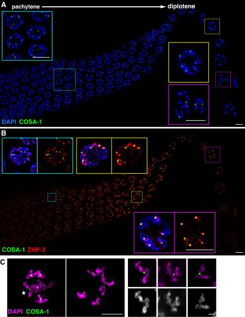
Crossovers (COs) between homologous chromosomes ensure their faithful segregation during meiosis. We identify C. elegans COSA-1, a cyclin-related protein conserved in metazoa, as a key component required to convert meiotic double-strand breaks (DSBs) into COs. During late meiotic prophase, COSA-1 localizes to foci that correspond to the single CO site on each homolog pair and indicate sites of eventual concentration of other conserved CO proteins. Chromosomes gain and lose competence to load CO proteins during meiotic progression, with competence to load COSA-1 requiring prior licensing. Our data further suggest a self-reinforcing mechanism maintaining CO designation. Modeling of a nonlinear dose-response relationship between IR-induced DSBs and COSA-1 foci reveals efficient conversion of DSBs into COs when DSBs are limiting and a robust capacity to limit cytologically differentiated CO sites when DSBs are in excess. COSA-1 foci serve as a unique live cell readout for investigating CO formation and CO interference.


Figures

Figure 1
Figure 1. COSA-1 is required to convert meiotic DSBs into interhomolog COs
(A) Full karyotypes of individual diakinesis-stage oocytes. 6 DAPI-stained bodies in the wild-type nucleus correspond to 6 pairs of homologs connected by chiasmata; 12 individual chromosomes (univalents) in the cosa-1 mutant nucleus reflect a lack of chiasmata. (B) cosa-1mutant pachytene nuclei in which pairing was assessed either by FISH at the 5S rDNA locus (chromosome V) or by immunostaining for X-chromosome pairing center (X-PC) binding protein HIM-8. A single FISH or HIM-8 signal in each nucleus indicates successful pairing. (C) cosa-1 mutant pachytene nuclei, showing colocalization of SC lateral element protein HIM-3 and SC central region protein SYP-1 between parallel tracks of DAPI-stained chromatin. (D) Immunolocalization of RAD-51 in mid to late pachytene nuclei in the cosa-1 mutant. RAD-51 foci indicative of DSB formation are abundant in mid-pachytene, and are greatly reduced or absent in most late pachytene nuclei (asterisk indicates an apoptotic nucleus). (E) Early diplotene nuclei. In WT, SYP-1 and HTP-1/2 are localized to reciprocal domains on each chromosome pair; in cosa-, this indicator of CO formation is not observed, as SYP-1 and HTP-1/2 remain extensively colocalized. Scale bar = 5 μm.
Figure 2
Figure 2. COSA-1 is a distant member of the cyclin superfamily with orthologs in metazoa
(A) Top, predicted gene structure of C. elegans cosa-l with mutant alleles indicated. Gray, UTR; magenta, coding exons. Bottom, construct used to express GFP::COSA-1. Green, GFP coding sequence; blue, extra tags and linker sequences. (B) Phylogenetic tree depicting a sampling of metazoan species. Green indicates lineages where COSA-1, MSH-4 and MSH-5 orthologs are all present; red indicates the absence of all three from Drosophilid species. (C) Predicted structure of residues 56-360 of C. elegans COSA-1 (yellow and magenta) aligned with crystal structure of residues 167-426 of human cyclin B1 (cyan). N-terminal residues of COSA-1 and cyclin B1 were removed to aid visualization of the two core cyclin fold motifs. In canonical cyclins, cyclin-fold motifs consist of 5 α-helices, with well-conserved interhelical angles in the N-terminal cyclin box motif. In the predicted COSA-1 structure, the N-terminal cyclin box is interrupted by an insertion of 33 amino acids, modeled here as an extension of α-helix 2 and an additional helix (magenta, α-2.5). Predicted α-helices 3-5 of COSA-1 align well with the corresponding helices of cyclin B1 and cyclin A, which contribute to the cyclin/CDK interface in Cyclin A/CDK2 (Jeffrey et al 1995).
Figure 3
Figure 3. GFP::COSA-1 localizes to foci corresponding to CO sites
(A and B) IF images of a portion of a gonad extending from mid-pachytene through diplotene and early diakinesis. GFP::COSA-1 foci are detected from late pachytene through early diakinesis. (A) Left inset: late pachytene nuclei, each containing 6 bright foci. Right insets: diplotene nuclei, with one focus on each chromosome pair; bottom panel shows COSA-1 foci positioned at the site of the single emerging chiasma on each chromosome pair. (B) Relationship between localization of GFP::COSA-1 and ZHP-3. Top left inset: 6 COSA-1 foci in a mid-pachytene nucleus with ZHP-3 in long stretches along the chromosomes. Top right inset: COSA-1 localized at one end of each comet-like stretch of ZHP-3. Bottom inset: COSA-1 and ZHP-3 colocalization in a diplotene nucleus. (C) Representative images of GFP::COSA-1 localization in late diplotene/early diakinesis nuclei, highlighting the location of GFP::COSA-1 at the site of the single emerging chiasma on each chromosome pair. Large panels, full projections of entire nuclei showing all 6 bivalents; asterisk indicates a bivalent depicted in smaller panels, which shows partial projections of individual bivalents. Scale bars = 5 μm except in the single bivalent panels in (C), where scale bar = 1 μm.
Figure 4
Figure 4. MSH-5 colocalizes with and depends on COSA-1
(A) IF images showing that MSH-5 foci are detected in mid-pachytene nuclei in excess of eventual COs (cyan inset), then decline by late pachytene, when they co-localize with GFP::COSA-1 foci (yellow inset). (B) Left, late pachytene nuclei from a wild-type germ line, showing comet-like localization of ZHP-3 with COSA- foci at the comet heads. Right, late pachytene nuclei from a cosa-1 mutant, showing persistence of ZHP-3 localization along the length of the chromosomes and a lack of MSH-5 foci. Scale bars = 5 μm; for insets, scale = 1 μm.
Figure 5
Figure 5. Time course of localization of CO proteins at IR-induced recombination sites
Immunolocalization of CO proteins (GFP::COSA-1, MSH-5 and/or ZHP-3) in pachytene nuclei from gfp::cosa-1; spo-11 worms, either in the absence of IR (A, left) or at the indicated times following exposure to 1 kRad IR. Scale bars = 5 μm. (A) Localization of COSA-1 and ZHP-3 or MSH-5 in late pachytene nuclei in the absence of IR (pre-IR) and 8 hr post-IR. In the unirradiated spo-11 control, ZHP-3 persists along the lengths of the chromosomes and the majority of nuclei lack COSA-1 and MSH-5 foci; a subset of nuclei have one or two COSA-1/MSH-5 aggregates (indicated by asterisks). 8 hr post-IR: 6 bright COSA-1 foci localize at the heads of comet-like ZHP-3 signals. (B) Mid to late pachytene region of a 1 hr post-IR germ line. Abundant IR-induced MSH-5 foci are detected specifically in mid-pachytene nuclei (left), while MSH-5 foci are not detected above baseline in late pachytene nuclei (right; 0, 1 or 2 MSH-5 signals colocalize with COSA-1, as in unirradiated controls). (C) GFP::COSA-1 localization in nuclei within the late pachytene region at 2.5 and 4 hrs post-IR; fields also include a few mid-pachytene nuclei (at the left) and a few early diplotene nuclei (at the right). Circles indicate nuclei in which 6 COSA-1 foci are detected. At 2.5 hrs post-IR, nuclei with 6 COSA-1 foci are limited to a narrow zone near the start of the late pachytene region. At 4hrs post-IR, the zone of nuclei with 6 COSA-1 foci has expanded, presumably reflecting movement into and progression through late pachytene of nuclei that had been exposed to IR during mid-pachytene. (D) Localization of MSH-5 and COSA-1 at 8 hrs post-IR in a region spanning the mid-to-late pachytene transition. Left inset: Mid-pachytene nuclei, showing MSH-5-only foci, in excess of eventual COs. Right inset: Late pachytene nuclei, showing 6 MSH-5 foci that co-localize with 6 COSA-1 foci.
Figure 6
Figure 6. Dose-response analysis reveals a highly non-linear relationship between IR-induced DSBs and COSA-1 foci
(A) Paired IF images showing GFP::COSA-1 foci in late pachytene nuclei from gfp::cosa-1; spo-11 germ lines exposed to the indicated IR doses, fixed 8 hrs post-IR, with numbers of foci in each nucleus indicated. Scale bar = 5 μm. (B) Stacked bar graph showing percentages of nuclei with the indicated numbers of COSA-1 foci at different IR doses. (C) Graph showing the highly non-linear relationship between IR dose and the mean number of COSA-1 foci per nucleus. Experimental data points are plotted in red, with error bars indicating standard deviation. Our mathematical model (μ = 6(1-e-cr) is plotted in blue; see Results and Supplemental Analysis. (D) Graph depicting linear relationship between IR dose and inferred mean number of DSBs per chromosome pair, calculated from our empirical data based on the postulates of our model. Empirical data points are in red, with linear regression in blue.
Figure 7
Figure 7. Relationship of COSA-1 to COs in conditions that alter CO number
(A) Graph showing percentages of nuclei with the indicated numbers of COSA-1 foci in strains with altered numbers of COs. Numbers of foci in the rtel-1(tm1866) and dpy-28(s939) mutants did not differ significantly from the control (Mann-Whitney test). Worms homozygous for the mnT12(X;IV) fusion chromosome have only 5 chromosome pairs, and mnT12 undergoes only one CO in the majority of meioses; an average of 5.3 COSA-1 foci per nucleus were observed in mnT12 homozygotes. Numbers of COSA-1 foci in rtel-1; mnT12 worms did not differ significantly from mnT12 controls. *WT control contains the gfp::cosa-1 transgene in an otherwise wild-type background. (B) The rtel-1 mutation does not suppress the lack of chiasmata caused by loss of cosa-1 function. Graph shows percent of diakinesis nuclei with a given number of DAPI-stained bodies. As in cosa-1(tm3298) single mutants, 11-12 DAPI bodies were detected in rtel-1; cosa-1 double mutants, reflecting a lack of chiasmata. Numbers of oocyte nuclei scored: wild type (n = 164), rtel-1 (n = 125), cosa-1 (n = 116), rtel-1; cosa-1 (n = 114). (C) Bar graph indicating genetic map distances (cM ± 95% C.I.) for the unc-60 dpy-11 interval measured for worms of the indicated genotypes (see Supplemental Experimental Procedures). ** indicates p < 0.001; * indicates p = 0.01. The CO frequency in the rtel-1 mutant (19.6 cM) was significantly elevated over wild type (13.4 cM, p = 0.0002; Fisher exact) and cosa-1/ + (13.6 cM, p = 0.0006) controls, which did not differ from each other. rtel-1 also differed significantly from rtel-1; cosa-1/+ (15.6 cM, p = 0.01), indicating that elevation in CO frequency was suppressed in rtel-1; cosa-1/+ worms. (D) Paired 3D volume renderings of a representative nucleus used to quantify X chromosome associated COSA-1 foci in the dpy-28 mutant. Staining for chromosome axis protein HTP-3 reveals the paths of synapsed chromosome pairs; arrow indicates the X chromosome, marked by X-PC associated protein HIM-8. Scale bar = 2 μm. Expected incidence of X chromosomes with 2 or more COSA-1 foci was estimated to be ≥ 36% based on frequencies of 2-CO and 3-CO products detected by genetic assay (Tsai et al., 2008).

Comment in

References

    1. Agarwal S, Roeder GS. Zip3 provides a link between recombination enzymes and synaptonemal complex proteins. Cell. 2000;102:245–255. - PubMed
    1. Anderson LK, Reeves A, Webb LM, Ashley T. Distribution of crossing over on mouse synaptonemal complexes using immunofluorescent localization of MLH1 protein. Genetics. 1999;151:1569–1579. - PMC - PubMed
    1. Barber LJ, Youds JL, Ward JD, McIlwraith MJ, O'Neil NJ, Petalcorin MI, Martin JS, Collis SJ, Cantor SB, Auclair M, et al. RTEL1 maintains genomic stability by suppressing homologous recombination. Cell. 2008;135:261–271. - PMC - PubMed
    1. Baudat F, de Massy B. Regulating double-stranded DNA break repair towards crossover or non-crossover during mammalian meiosis. Chromosome Res. 2007;15:565–577. - PubMed
    1. Bhalla N, Wynne DJ, Jantsch V, Dernburg AF. ZHP-3 acts at crossovers to couple meiotic recombination with synaptonemal complex disassembly and bivalent formation in C. elegans. PLoS Genet. 2008;4:e1000235. - PMC - PubMed

Publication types

LinkOut - more resources