

Automatic motion compensation of free breathing acquired myocardial perfusion data by using independent component analysis
- PMID: 22465078
- PMCID: PMC3372575
- DOI: 10.1016/j.media.2012.02.004
Automatic motion compensation of free breathing acquired myocardial perfusion data by using independent component analysis
Abstract
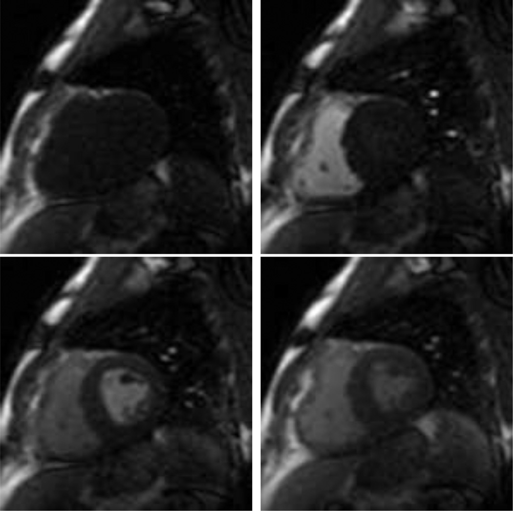
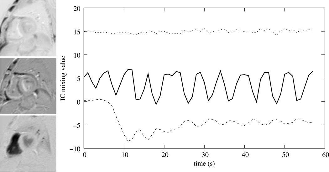
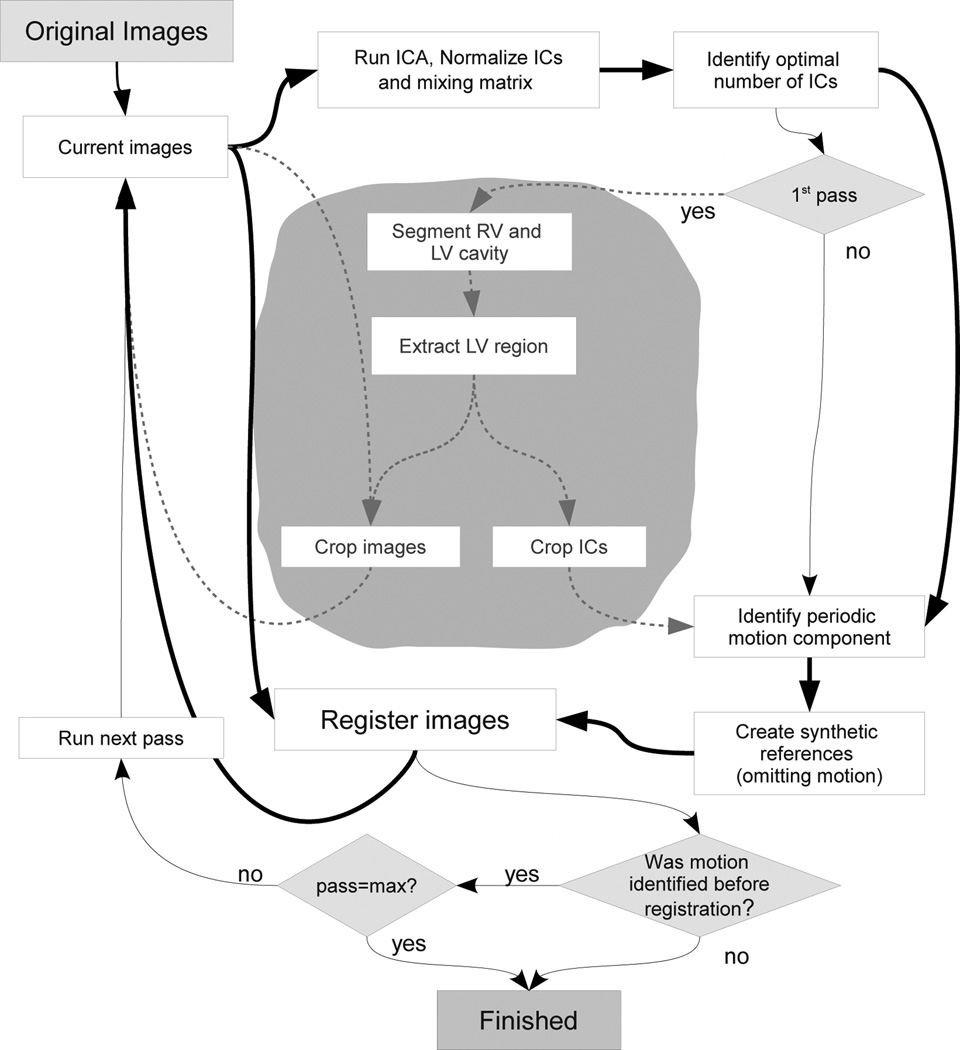
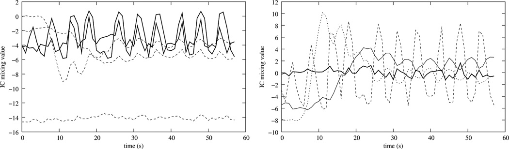
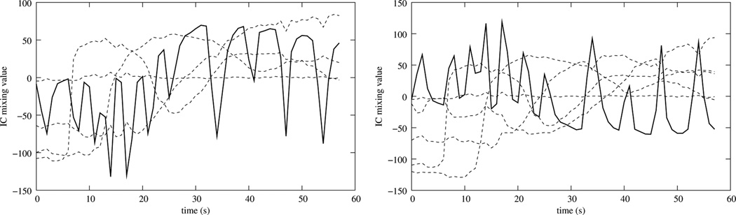
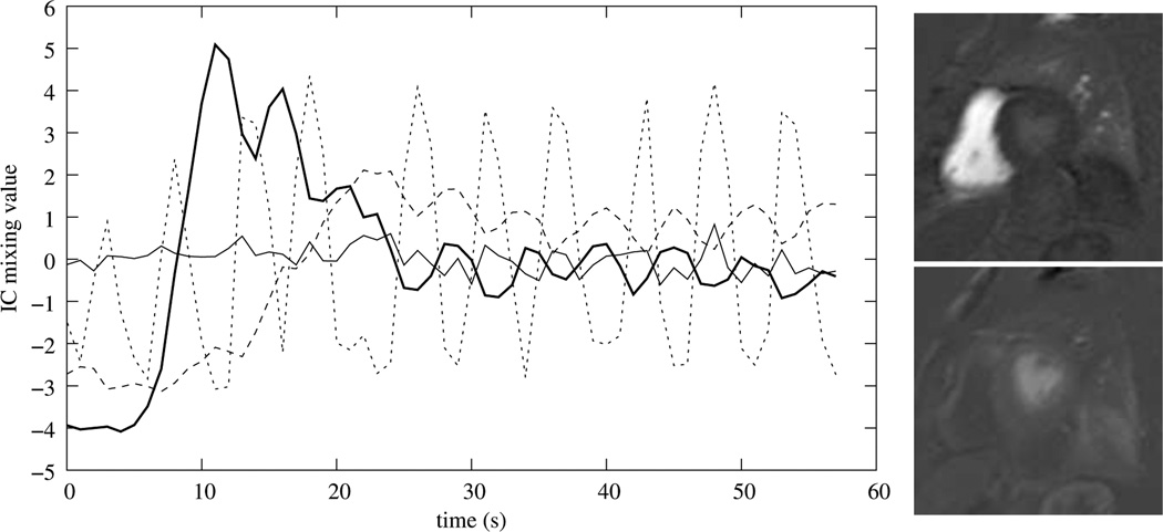
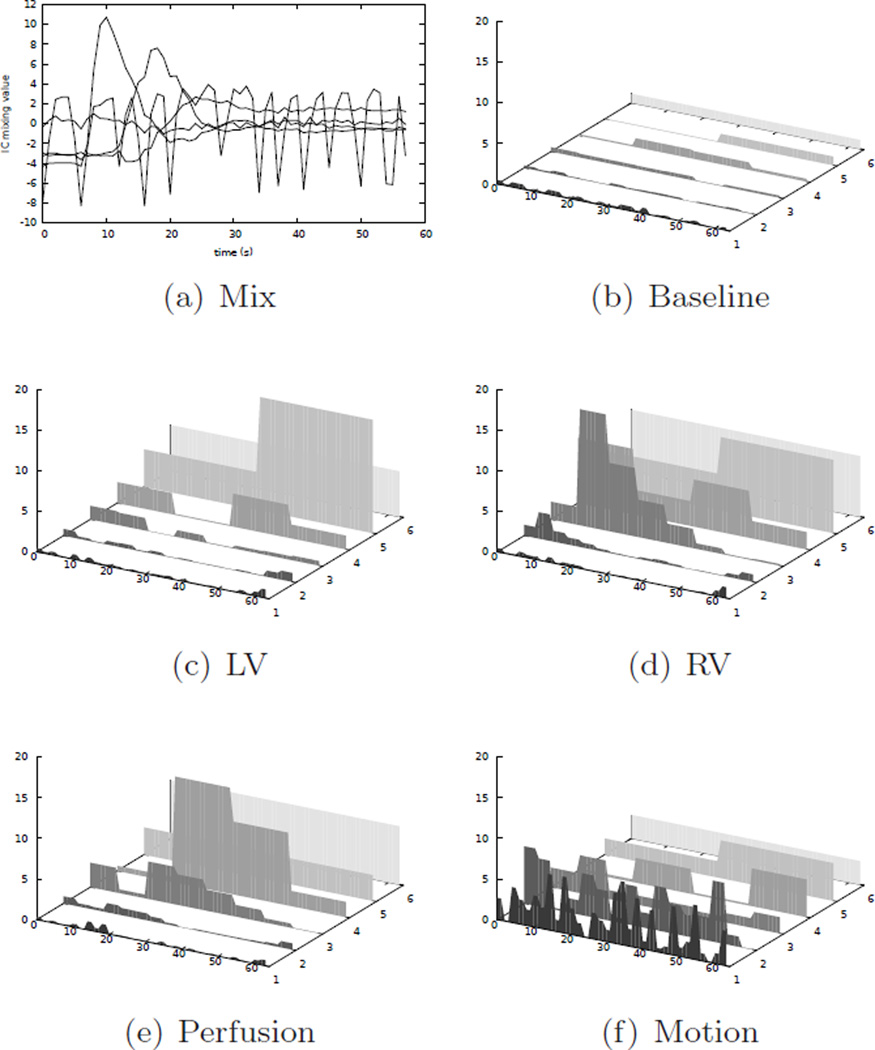
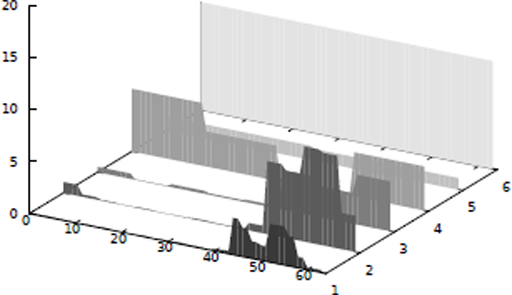
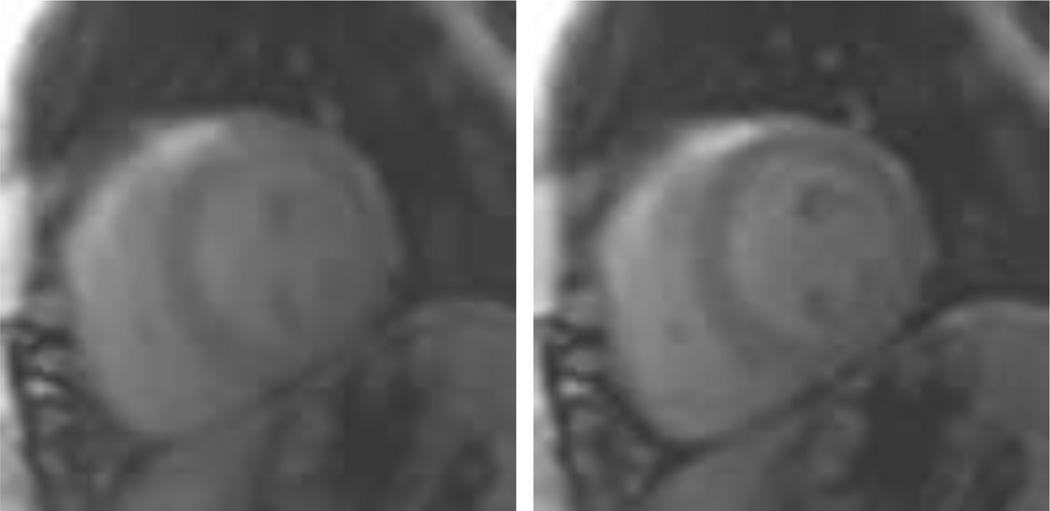
Images acquired during free breathing using first-pass gadolinium-enhanced myocardial perfusion magnetic resonance imaging (MRI) exhibit a quasiperiodic motion pattern that needs to be compensated for if a further automatic analysis of the perfusion is to be executed. In this work, we present a method to compensate this movement by combining independent component analysis (ICA) and image registration: First, we use ICA and a time-frequency analysis to identify the motion and separate it from the intensity change induced by the contrast agent. Then, synthetic reference images are created by recombining all the independent components but the one related to the motion. Therefore, the resulting image series does not exhibit motion and its images have intensities similar to those of their original counterparts. Motion compensation is then achieved by using a multi-pass image registration procedure. We tested our method on 39 image series acquired from 13 patients, covering the basal, mid and apical areas of the left heart ventricle and consisting of 58 perfusion images each. We validated our method by comparing manually tracked intensity profiles of the myocardial sections to automatically generated ones before and after registration of 13 patient data sets (39 distinct slices). We compared linear, non-linear, and combined ICA based registration approaches and previously published motion compensation schemes. Considering run-time and accuracy, a two-step ICA based motion compensation scheme that first optimizes a translation and then for non-linear transformation performed best and achieves registration of the whole series in 32±12s on a recent workstation. The proposed scheme improves the Pearsons correlation coefficient between manually and automatically obtained time-intensity curves from .84±.19 before registration to .96±.06 after registration.
Copyright © 2012 Elsevier B.V. All rights reserved.
Figures


References
-
- Breeuwer M, Spreeuwers L, Quist M. Automatic quantitative analysis of cardiac MR perfusion images. Proceedings of the SPIE - The International Society for Optical Engineering. 2001:733–742.
-
- Comon P. Independent Component Analysis, a new concept? Signal Processing, Special issue on Higher-Order Statistics. 1994;36:287–314.
-
- Daubechies I. Orthonormal Bases of Compactly Supported Wavelets. Communications on Pure and Applied Mathematics. 1988;41 909996.
-
- Delzescaux T, Frouin F, Cesare AD, Philipp-Foliguet S, Todd-Pokropek A, Herment A, Janier M. Using an adaptive semiautomated self-evaluated registration technique to analyze mri data for myocardial perfusion assessment. Journal of Magn. Reson. Imag. 2003;18:681–690. - PubMed
-
- Dornier C, Ivancevic MK, Thevenaz P, Vallee JP. Improvement in the quantification of myocardial perfusion using an automatic splinebased registration algorithm. Journal of Magn. Reson. Imag. 2003;18:160–168. - PubMed
Publication types
MeSH terms
Grants and funding
LinkOut - more resources
Full Text Sources
Medical
Miscellaneous
