. 2012 Apr 20;420(4):816-21.
doi: 10.1016/j.bbrc.2012.03.080. Epub 2012 Mar 23.

Chlamydia trachomatis Tarp cooperates with the Arp2/3 complex to increase the rate of actin polymerization

Affiliations

Chlamydia trachomatis Tarp cooperates with the Arp2/3 complex to increase the rate of actin polymerization

Shahanawaz Jiwani et al. Biochem Biophys Res Commun. .

Abstract

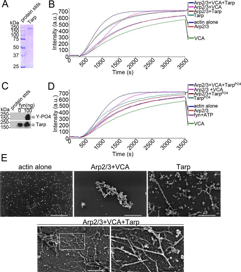
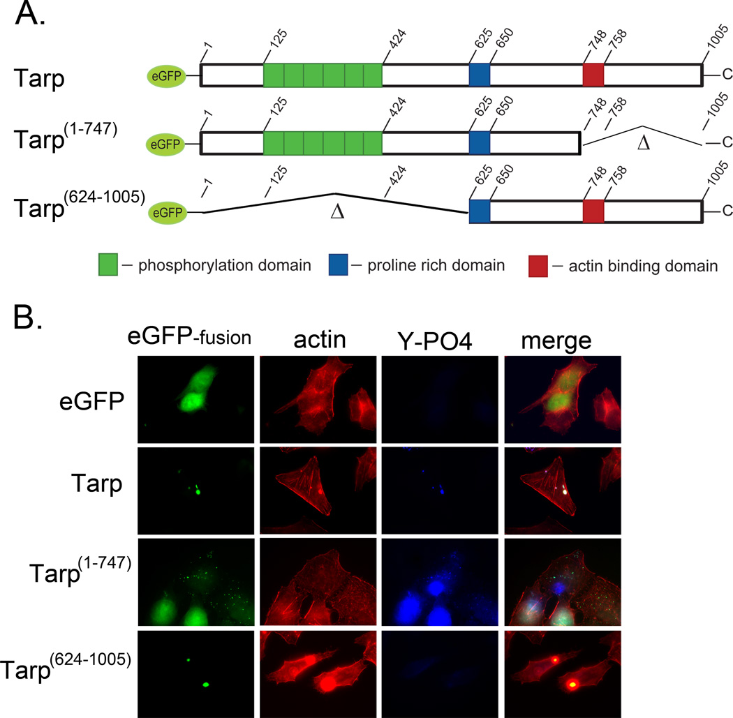
Actin polymerization is required for Chlamydia trachomatis entry into nonphagocytic host cells. Host and chlamydial actin nucleators are essential for internalization of chlamydiae by eukaryotic cells. The host cell Arp2/3 complex and the chlamydial translocated actin recruiting phosphoprotein (Tarp) are both required for entry. Tarp and the Arp2/3 complex exhibit unique actin polymerization kinetics individually, but the molecular details of how these two actin nucleators cooperate to promote bacterial entry is not understood. In this study we provide biochemical evidence that the two actin nucleators act synergistically by co-opting the unique attributes of each to enhance the dynamics of actin filament formation. This process is independent of Tarp phosphorylation. We further demonstrate that Tarp colocalization with actin filaments is independent of the Tarp phosphorylation domain. The results are consistent with a model in which chlamydial and host cell actin nucleators cooperate to increase the rate of actin filament formation.


Figures

Fig. 1
Fig. 1
Tarp and Arp2/3 complex cooperate to polymerize actin. (A) Purified recombinant Tarp employed for these studies was resolved by SDS-PAGE and visualized by Coomassie blue staining. (B) Pyrene actin polymerization in the presence of Tarp and activated Arp2/3 complex (Arp2/3+VCA+Tarp) was compare to actin polymerization kinetics initiated by Tarp alone (Tarp), activated Arp2/3 complex alone (Arp2/3+VCA) and Tarp with an inactive Arp2/3 complex (Arp2/3+Tarp). Actin alone (actin), inactive Arp2/3 complex (Arp2/3) and the Verprolin, Cofilin, Acidic (VCA) domain of N-WASP served as additional controls. Bacterial and host cell nucleators were incubated with pyrene conjugated actin and actin polymerization was measured by an increase in fluorescence of pyrenyl-actin incorporated into actin filaments (Intensity (a.u.)) over approximately 1 hour (Time (s)) following the addition of polymerization buffer at 300 seconds. (C) Purified recombinant Tarp was phosphorylated by the Src family kinase member fyn. Tarp proteins were incubated with 100ng of purified fyn kinase and ATP and following a short incubation were suspended in protein sample buffer and resolved by SDS-PAGE followed by transfer to nitrocellulose. Immunoblots were performed with Tarp (α Tarp) and phosphotyrosine (α Y-PO4) specific antisera. (D) Pyrene actin polymerization assays as B but with phosphorylated Tarp. Additional controls include actin polymerization assays performed in the presence of fyn kinase and phosphorylation buffer containing ATP (fyn+ATP)(E) Electron micrographs of actin filaments nucleated by Tarp and the Arp2/3 complex. Protein samples were removed from actin polymerization experiments similar to those shown in B at 900 seconds (10 minutes after the addition of polymerization buffer) and actin filaments were visualized by scanning electron microscopy. White scale bar represents 400 nm and black scale bar represents 150 nm.
Fig. 2
Fig. 2
Actin filaments co-localize with ectopically expressed Tarp in a phosphorylation domain independent manner. Tarp proteins ectopically expressed as enhanced green fluorescence protein fusions were examined for their ability to localize with actin filaments. (A) Schematic of the eGFP-Tarp fusion proteins indicating the location of the actin binding domain (red box), the proline rich domain (blue box) and the tyrosine rich phosphorylation domain (green boxes). Δ indicates amino acids deleted in the mutant eGFP –Tarp fusion proteins and numbers indicate amino acid positions encoded within the C. trachomatis tarP gene (B) Host cells expressing eGFP (eGFP) alone or eGFP fusions (eGFP-fusion) of full length Tarp (Tarp), or deletion mutants lacking the phosphorylation domain (Tarp 624–1005) or actin binding domain (Tarp 1–747) but harboring the proline rich domain (PRD) required for protein aggregation were fixed and stained with Alexa fluor 568 conjugated phalloidin (actin), phosphotyrosine specific antisera (Y-PO4) and goat anti-mouse conjugated to Alexa 350.

References

    1. CDC Grand Rounds: Chlamydia prevention: challenges and strategies for reducing disease burden and sequelae. MMWR Morb Mortal Wkly Rep. 60:370–373. - PubMed
    1. Schachter J. Infection and disease epidemiology. In: Stephens RS, editor. Chlamydia; Intracellular biology, pathogenesis, and immunity. Washington, D.C.: ASM Press; 1999. pp. 139–169.
    1. Mariotti SP, Pascolini D, Rose-Nussbaumer J. Trachoma: global magnitude of a preventable cause of blindness. Br J Ophthalmol. 2009;93:563–568. - PubMed
    1. Hybiske K, Stephens RS. Mechanisms of host cell exit by the intracellular bacterium Chlamydia. Proc Natl Acad Sci U S A. 2007;104:11430–11435. - PMC - PubMed
    1. Moulder JW. Interaction of chlamydiae and host cells in vitro. Microbiol Rev. 1991;55:143–190. - PMC - PubMed

Publication types

MeSH terms

LinkOut - more resources