. 2012 Jun;92(3):318-26.
doi: 10.1016/j.yexmp.2012.02.003. Epub 2012 Mar 21.

Mallory-Denk bodies form when EZH2/H3K27me3 fails to methylate DNA in the nuclei of human and mice liver cells

Affiliations

Mallory-Denk bodies form when EZH2/H3K27me3 fails to methylate DNA in the nuclei of human and mice liver cells

B A French et al. Exp Mol Pathol. 2012 Jun.

Abstract

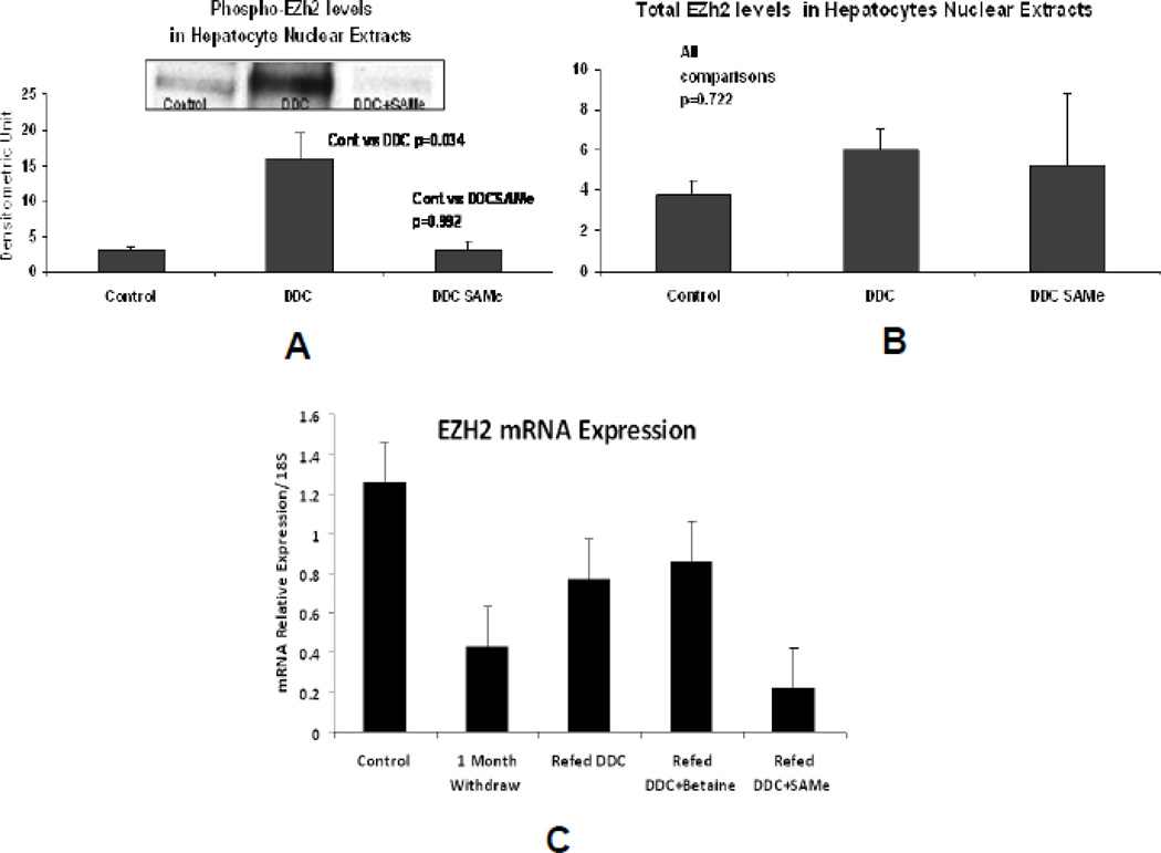
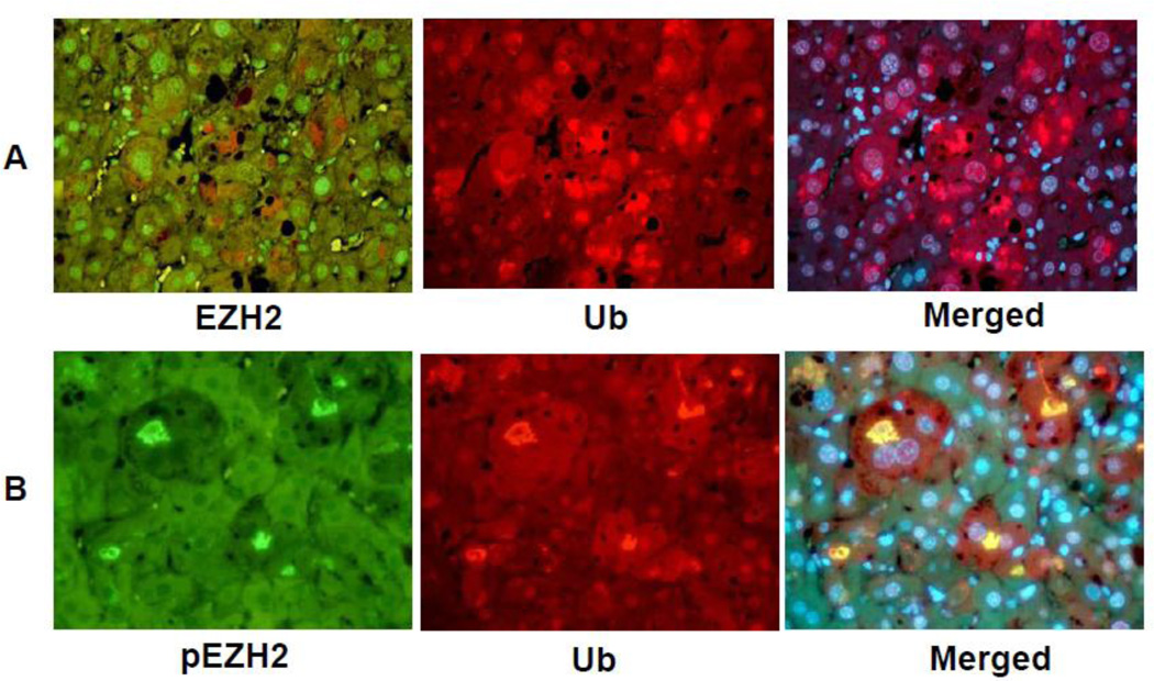
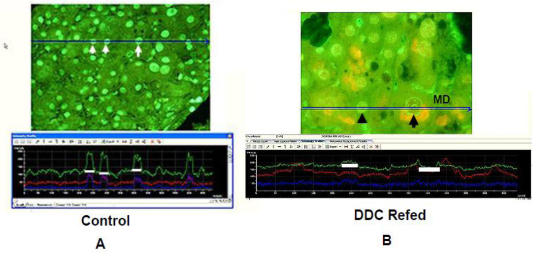
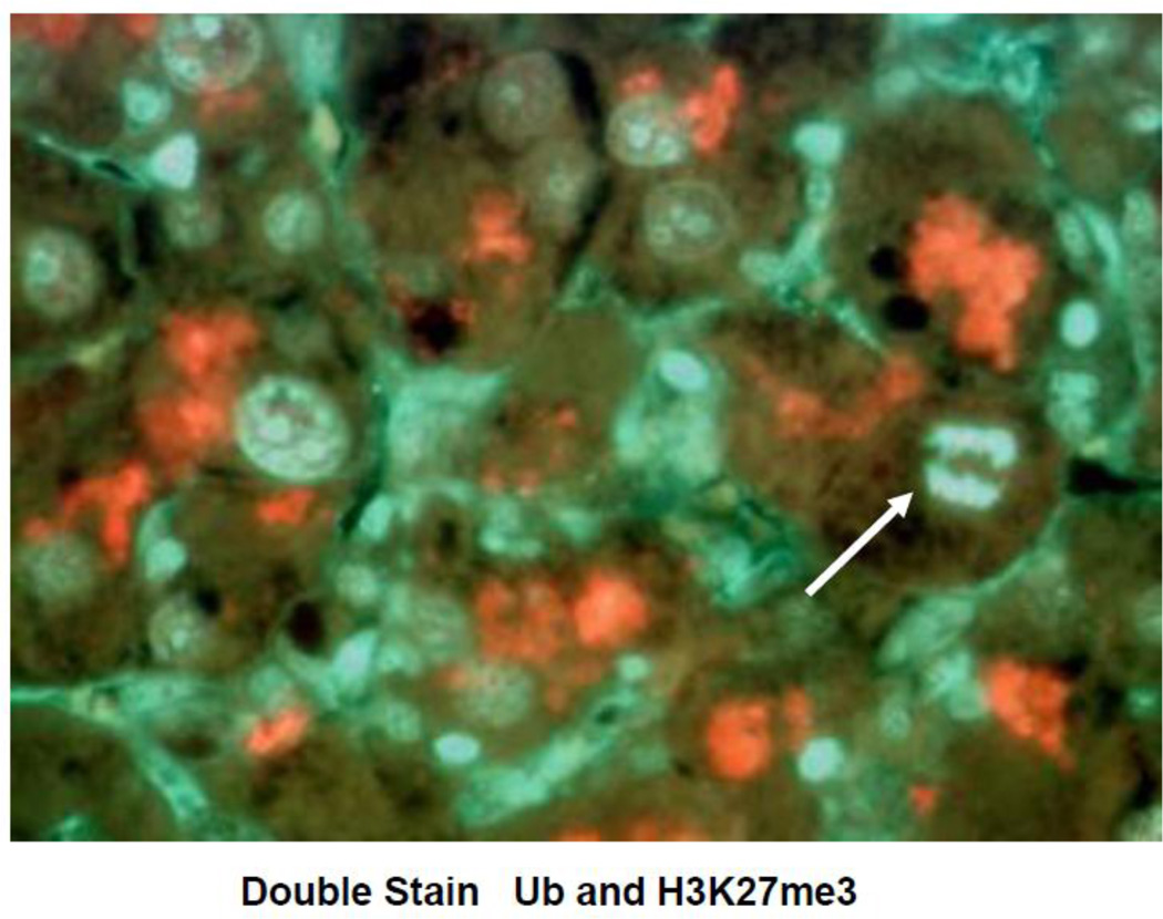
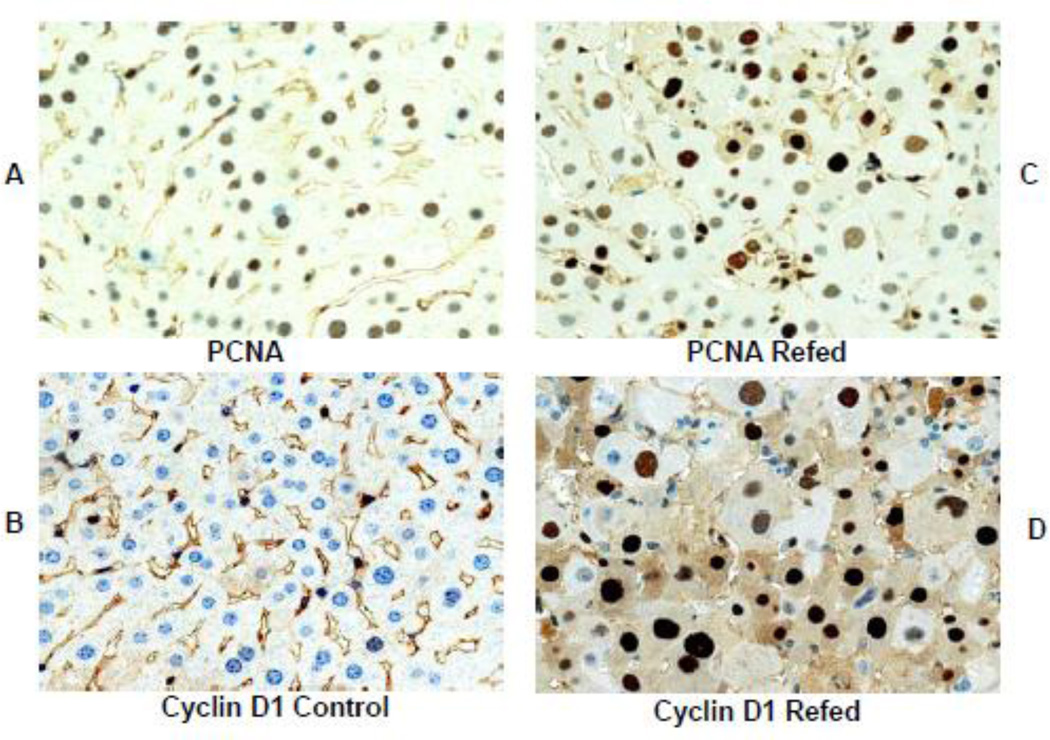
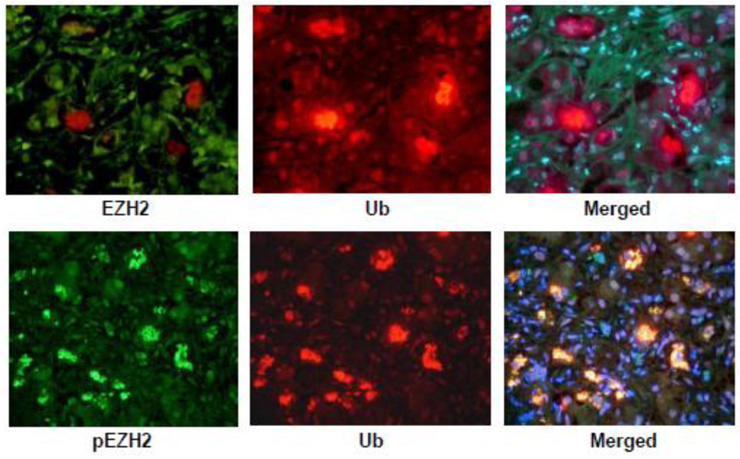
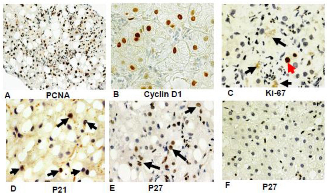
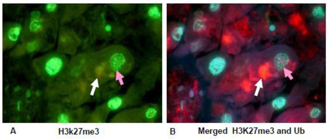
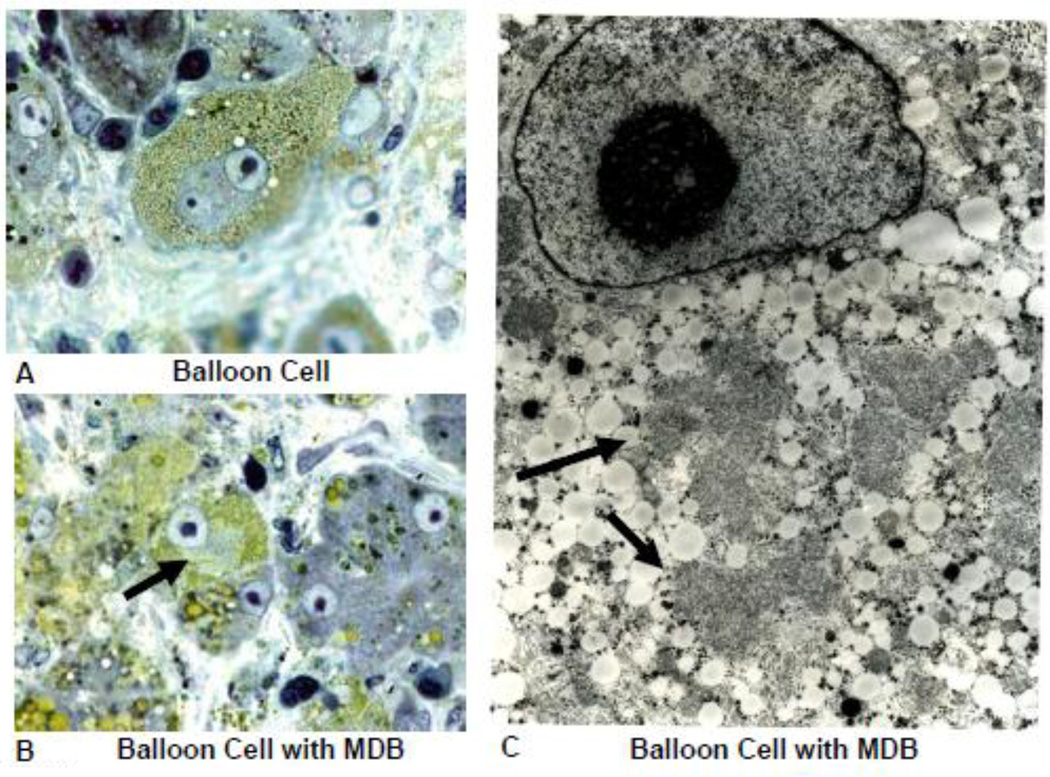
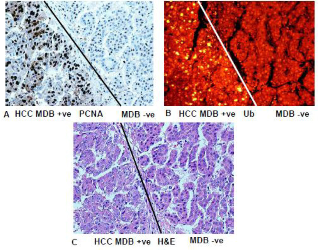
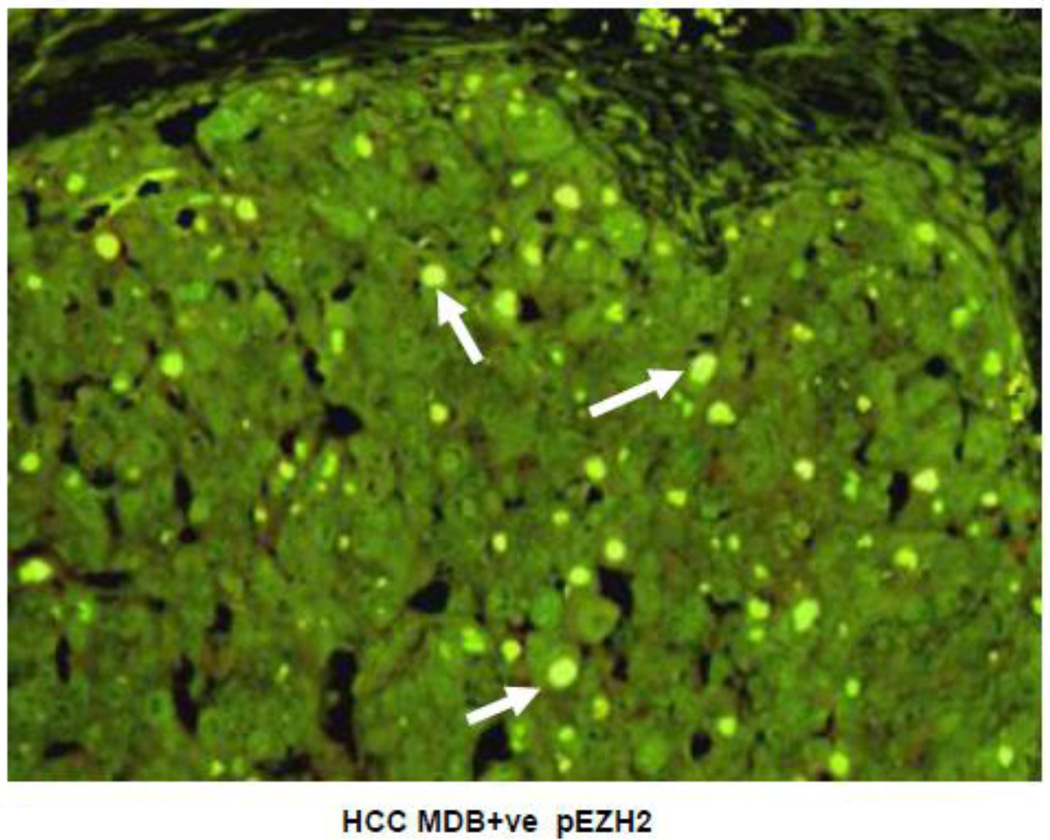
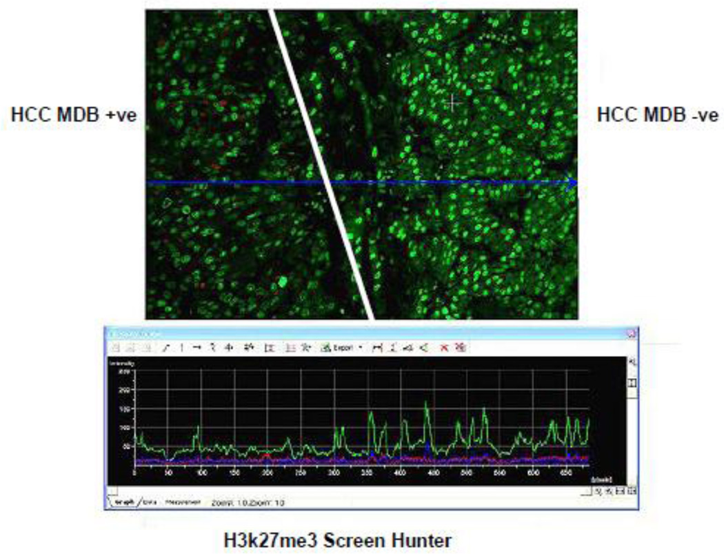
EZH2/H3K27me3 and polycomb group complex (PcG) play a major role in regulating global gene expression including tumor suppressor genes. EZH2 is linked to cell cycle regulated EZH2 phosphorylation by CDK1, a mitotic kinase which increases in arrested mitosis compared to S phase. CDK1 phosphorylation of EZH2 accelerates the degradation of pEZH2. Phospho-EZH2 is subjected to ubiquitination. The half-like of pEZH2 is shorter when compared to total EZH2. In the present study, pEZH2 was found concentrated together with ubiquitin in the Mallory-Denk bodies (MDB) that were formed in hepatocytes in the livers of drug primed mice refed DDC and humans with alcoholic hepatitis or hepatocellular carcinoma. The cells that formed MDBs in the mice livers studied were associated with a growth advantage and a high proliferative index. However, the livers from patients with alcoholic hepatitis showed evidence of cell cycle arrest where PCNA, cyclin D1 and p27 positive nuclei were numerous but Ki-67 positive nuclei were scarce. It is concluded that MDB formation is linked to the cell cycle and global gene expression (i.e. loss of gene silencing) through its association with the regulation of the polycomb group PRC2/EZH2/H3K27me3 complex.


Figures

Fig 1
Fig 1
A) Western blot using the antibody to pEZH2 showed an increase in proteins in the n mean±SEM). B) Western blots using the antibody to EZH2 showed there was no significant difference between the 3 groups of mice used in A (n=3). C) qPCR of the nuclear levels of EZH2 mRNA showed no significant difference in the levels between the controls and the DDC refed groups of mice except when SAMe was fed the DDC refed mice (n=3, mean±S.E.M., p<0.05).
Fig 2
Fig 2
Liver cells which formed MDBs stained positive for ubiquitin (A and B, red) and pEZH2 (A, green) but not EZH2 (B, green). pEZH2 and ubiquitin were co localized in MDBs (B, yellow). (A×479, B×572.)
Fig 3
Fig 3
The Mallory-Denk body (MDB) hepatocytic balloon cell is shown in the upper panel. The stain for H3K27me3 in the nucleus of the balloon cell is less intense than the adjacent normal hepatocytic nucleus (arrow) and the nuclei in the control liver in the lower panel. The balloon cell MDB stains positive for ubiquitin (upper panel). The merged photo of the balloon cell further documents the reduced nuclear staining for H3/K27me3 (Confocal microscope).
Fig 4
Fig 4
The MDB forming hepatocytic balloon cell nucleus in the liver of a DDC refed mouse shows the loss of staining intensity (green) for H3K27me3 (B) when compared to the neighboring normal hepatocytes. The normal control (A) shows equal intensity staining of the nuclei for H3K27me3, (B) (arrow) MDBs stain yellow because H3K27me3 (green) is double stained with ubiquitin (red). The reduction in nuclear levels of H3K27me3 induced by DDC refeeding has been confirmed by Western blot (Bardag-Gorce et al., 2010). (Screen Hunter) (A×543, B×654)
Fig 5
Fig 5
Liver from a DDC refed mouse double stained with an antibody to ubiquitin and DAPI (blue) shows that the nucleus of a ballooned hepatocyte that had formed an MDB was in mitoses (arrow) (x 1962).
Fig 6
Fig 6
Sections of liver from mice refed DDC and control stained with an antibody to PCNA showing rare nuclear staining positive in the control (A) and numerous positive staining in the DDC refed mouse liver (C). Similarly liver from the mouse refed DDC stained for cyclin D1 (D) shows numerous positive stained nuclei. The control mouse liver shows only a few positive stained nuclei. Magnifications are; (A×606), (B×606), (C×606), (D×910).
Fig 7
Fig 7
A liver biopsy from a patient who had alcoholic hepatitis was stained with an antibody to EZH2 (upper panel) and an antibody to pEZH2 (lower panel). Note that the nuclei stained for EZH2 were all stained with the same intensity (left photo). The MDBs were stained green for pEZH2, red for ubiquitin and yellow when the tricolor filter was used indicating that pEZH2 was increased in MDBs. pEZH2 was also increased in the Western blot in mice refed DDC (Fig 1) and the liver section stained with the pEZH2 antibody (Fig 2). (Upper and Lower Panel×436).
Fig 8
Fig 8
The liver biopsy slides from patients with alcoholic hepatitis were stained with antibodies to PCNA (A), Cyclin D1 (B), Ki-67 (C), p21 (D) and p27 (E). Only a few scattered nuclei were positive for Ki67 (arrow). No nuclei stained positive in a liver from an alcoholic hepatitis patient. (F). Magnification (A×218), (B×654), (C×654), (D×684), (E×436), F×436).
Fig 9
Fig 9
Liver section from a patient who died with alcoholic hepatitis, stained for H3K27me3 and ubiquitin. Note that the liver cell containing an MDB (white arrow, red) B) stained with less intensity for H3K27me3 (orange arrow) when compared to the nucleus of a neighboring hepatocyte. (A×1040), (B×1040).
Fig 10
Fig 10
Electron microscopic photograph of balloon cells from a patient with alcoholic hepatitis. A) Balloon cell with micro vesicular fat stained by osmium and a nucleus which contains a large nucleolus. B) Ballooning cell that formed an MDB (arrow). C) EM of balloon cell with micro vesicular fat in the cytoplasm together with an MDB (arrows). Note the nuclear euchromatin and prominent nucleolus, (A×1451), (B×1040), (C×6710).
Fig 11
Fig 11
Liver hepatocellular carcinoma has formed MDBs to the left of the line and not to the right of line in A, B, and C. A) PCNA stain shows that most of the nuclei in tumor cells that have formed MDBs stain positive. B) MDBs formed by the tumor cells stain positive for ubiquitin (yellow). C) Hematoxylin and eosin stain. The tumor cells that formed MDBs have vesicular nuclei compared to the tumor cells that have not formed MDBs on the right. (A×346), (B×346), (C×346).
Fig 12
Fig 12
Hepatocellular carcinoma double stained for pEZH2 and ubiquitin viewed with the tricolor filter showing co localization in MDBs (arrows). (x654).
Fig 13
Fig 13
Hepatocellular carcinoma, the same as shown in Fig 11 and 12, stained for H3K27me3. Note the tumor cell nuclei on the left that have formed MDBs stain with less intensity than the tumor cells on the right which have not formed MDBs. Note the green tracing of intensity below in the screen hunter (x 206).

References

    1. Abbas T, Dutta A. CRL4Cd+2; Master coordinator of cell cycle progression and genome stability. Cell cycle. 2011;10:241–249. - PMC - PubMed
    1. Abbas T, Dutta A. p21 in cancer intricate net works and multiple activities. Nat Rev Cancer. 2009;9:400–414. - PMC - PubMed
    1. Bardag-Gorce F, Dedes J, French BA, Oliva J, Li J, French SW. Mallory body formation is associated with epigenetic phenotypic change in hepatocytes in vivo. Exp Mol Pathol. 2007;83:160–168. - PMC - PubMed
    1. Bardag-Gorce F, Oliva J, Lin A, Li J, French BA, French SW. SAMe prevents the up regulation of toll-like receptor signaling in Mallory-Denk body forming hepatocytes. Exp Mol Pathol. 2010;88:376–379. - PMC - PubMed
    1. Bradford MM. A rapid and sensitive method for the quantitation of microgram quantities of protein utilizing the principle of protein-dye binding. Anal Biochem. 1976;72:248–254. - PubMed

Publication types

MeSH terms