. 2012 Jun 22;419(5):315-29.
doi: 10.1016/j.jmb.2012.03.014. Epub 2012 Mar 29.

Role of arginine 293 and glutamine 288 in communication between catalytic and allosteric sites in yeast ribonucleotide reductase

Affiliations

Role of arginine 293 and glutamine 288 in communication between catalytic and allosteric sites in yeast ribonucleotide reductase

Md Faiz Ahmad et al. J Mol Biol. .

Abstract

Ribonucleotide reductases (RRs) catalyze the rate-limiting step of de novo deoxynucleotide (dNTP) synthesis. Eukaryotic RRs consist of two proteins, RR1 (α) that contains the catalytic site and RR2 (β) that houses a diferric-tyrosyl radical essential for ribonucleoside diphosphate reduction. Biochemical analysis has been combined with isothermal titration calorimetry (ITC), X-ray crystallography and yeast genetics to elucidate the roles of two loop 2 mutations R293A and Q288A in Saccharomyces cerevisiae RR1 (ScRR1). These mutations, R293A and Q288A, cause lethality and severe S phase defects, respectively, in cells that use ScRR1 as the sole source of RR1 activity. Compared to the wild-type enzyme activity, R293A and Q288A mutants show 4% and 15%, respectively, for ADP reduction, whereas they are 20% and 23%, respectively, for CDP reduction. ITC data showed that R293A ScRR1 is unable to bind ADP and binds CDP with 2-fold lower affinity compared to wild-type ScRR1. With the Q288A ScRR1 mutant, there is a 6-fold loss of affinity for ADP binding and a 2-fold loss of affinity for CDP compared to the wild type. X-ray structures of R293A ScRR1 complexed with dGTP and AMPPNP-CDP [AMPPNP, adenosine 5-(β,γ-imido)triphosphate tetralithium salt] reveal that ADP is not bound at the catalytic site, and CDP binds farther from the catalytic site compared to wild type. Our in vivo functional analyses demonstrated that R293A cannot support mitotic growth, whereas Q288A can, albeit with a severe S phase defect. Taken together, our structure, activity, ITC and in vivo data reveal that the arginine 293 and glutamine 288 residues of ScRR1 are crucial in facilitating ADP and CDP substrate selection.


Figures

Fig. 1
Fig. 1
ITC profiles of effector (dGTP) and substrate (ADP or CDP) binding to wild-type, R293A and Q288A ScRR1. Effector and substrate binding was derived from the nonlinear least-squares fit of the isotherms. The isotherm profile of dGTP, ADP and CDP to the wild-type, R293A and Q288A ScRR1 could be best fitted to the one-site binding model. (a) Binding isotherm of dGTP to the wild-type ScRR1. (b) dGTP binding to the R293A ScRR1. (c and d) Binding isotherms of substrate ADP to dGTP-saturated wild-type and R293A ScRR1. (e and f) ITC profiles of substrate CDP binding to wild-type ScRR1 and R293A ScRR1. (g) Binding isotherm of dGTP to the Q288A ScRR1. (h) Binding isotherm of ADP to the Q288A ScRR1. (i) Binding isotherm of CDP to the Q288A ScRR1.
Fig. 1
Fig. 1
ITC profiles of effector (dGTP) and substrate (ADP or CDP) binding to wild-type, R293A and Q288A ScRR1. Effector and substrate binding was derived from the nonlinear least-squares fit of the isotherms. The isotherm profile of dGTP, ADP and CDP to the wild-type, R293A and Q288A ScRR1 could be best fitted to the one-site binding model. (a) Binding isotherm of dGTP to the wild-type ScRR1. (b) dGTP binding to the R293A ScRR1. (c and d) Binding isotherms of substrate ADP to dGTP-saturated wild-type and R293A ScRR1. (e and f) ITC profiles of substrate CDP binding to wild-type ScRR1 and R293A ScRR1. (g) Binding isotherm of dGTP to the Q288A ScRR1. (h) Binding isotherm of ADP to the Q288A ScRR1. (i) Binding isotherm of CDP to the Q288A ScRR1.
Fig. 2
Fig. 2
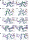
Stereo view of effectors and substrates (dGTP–ADP complex or AMPPNP–CDP) bound to wild-type, R293A and Q288A ScRR1. (a) |Fo| − |Fc| electron density for effector dGTP, loop 2 and substrate ADP of wild-type ScRR1 (blue density) contoured at 3σ. (b) |Fo| − |Fc| electron density for effector dGTP and loop 2 of R293A ScRR1 (blue density) contoured at 3σ. (c) |Fo| − |Fc| electron density for effector dGTP and loop 2 of Q288A ScRR1 (blue density) contoured at 3σ. (d) |Fo| − |Fc| electron density for effector AMPPNP, loop 2 and substrate CDP of wild-type ScRR1. (e) |Fo| − |Fc| electron density for effector AMPPNP, loop 2 and substrate CDP of R293A ScRR1 (blue density) contoured at 3σ. (f) |Fo| − |Fc| electron density for effector AMPPNP, disordered loop 2 and substrate CDP of Q288A ScRR1 (blue density) contoured at 3σ.
Fig. 2
Fig. 2
Stereo view of effectors and substrates (dGTP–ADP complex or AMPPNP–CDP) bound to wild-type, R293A and Q288A ScRR1. (a) |Fo| − |Fc| electron density for effector dGTP, loop 2 and substrate ADP of wild-type ScRR1 (blue density) contoured at 3σ. (b) |Fo| − |Fc| electron density for effector dGTP and loop 2 of R293A ScRR1 (blue density) contoured at 3σ. (c) |Fo| − |Fc| electron density for effector dGTP and loop 2 of Q288A ScRR1 (blue density) contoured at 3σ. (d) |Fo| − |Fc| electron density for effector AMPPNP, loop 2 and substrate CDP of wild-type ScRR1. (e) |Fo| − |Fc| electron density for effector AMPPNP, loop 2 and substrate CDP of R293A ScRR1 (blue density) contoured at 3σ. (f) |Fo| − |Fc| electron density for effector AMPPNP, disordered loop 2 and substrate CDP of Q288A ScRR1 (blue density) contoured at 3σ.
Fig. 3
Fig. 3
Comparison of AMPPNP- and CDP-bound wild-type and R293A ScRR1 structures. (a) Stereo view of the substrate CDP binding and its proximity to loop 2. (b) Comparison of catalytic site of wild-type and R293A ScRR1 with CDP bound. All catalytically important residues and their interactions with the wild-type ScRR1 CDP substrate are shown as broken lines. (c) Effect of the mutation on the loop 2 conformation. In all of the panels, wild-type ScRR1 is colored magenta and R293A ScRR1 is colored green and the interactions are shown as broken lines.
Fig. 3
Fig. 3
Comparison of AMPPNP- and CDP-bound wild-type and R293A ScRR1 structures. (a) Stereo view of the substrate CDP binding and its proximity to loop 2. (b) Comparison of catalytic site of wild-type and R293A ScRR1 with CDP bound. All catalytically important residues and their interactions with the wild-type ScRR1 CDP substrate are shown as broken lines. (c) Effect of the mutation on the loop 2 conformation. In all of the panels, wild-type ScRR1 is colored magenta and R293A ScRR1 is colored green and the interactions are shown as broken lines.
Fig. 4
Fig. 4
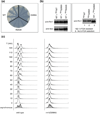
Growth defects of rnr1(R293A) and rnr1(Q288A) mutants. (a) R293A ScRR1 causes lethality. TRP1CEN plasmids harboring N-terminally 3×Myc-taggged wild-type RNR1 (WT), rnr1(Q288A) and rnr1(R293A) were introduced into a Δrnr1Δrnr3 double deletion strain that was kept alive by a copy of wild-type untagged RNR1 on a URA3CEN plasmid. Growth on 5-FOA-containing plates indicates loss of the URA3CENRNR1 plasmid and that the 3×Myc-tagged Rnr1 expressed from the TRP1CEN plasmid can provide the essential RNR1 activity. (b) In vivo expression of Q288A and R293A ScRR1 proteins. Left panel: Δrnr1Δrnr3 mutant cells harboring both the URA3CENRNR1 plasmid and the TRP1CEN plasmid encoding 3×Myc-tagged wild-type (MycWT), R293A or Q288A ScRR1 (MycR293A and MycQ288A) were grown to log phase and harvested for protein extraction and Western blotting with anti-Rnr1 and anti-Myc antibodies. Right panel: Western blotting detection of Rnr1 proteins using the polyclonal anti-Rnr1 antibodies in whole-cell extracts. Lanes 1 and 2 were from cells that grew on 5-FOA-containing plate. Only the 3×Myc-tagged Rnr1 (MycWTand MycQ288A) were detected by anti-Rnr1. Lane 3 is from cells without selection on the 5-FOA plate; both untagged wild-type (the lower band, WT) and 3×Myc-tagged (the upper band, MycR293A) Rnr1 proteins were detected. (c) Comparison of cell cycle progression of the wild-type and the rnr1(Q288A) mutant cells by flow cytometry. Δrnr1Δrnr3 double deletion strains harboring RNR1(WT) or rnr1(Q288A) were synchronized in G1 phase of the cell cycle by α-factor-mediated arrest and then released into cell cycle by washing off α factor. Cells were harvested at 10-min intervals at room temperature (23 °C) and processed for flow cytometry analysis. The shaded profiles are cells from asynchronously growing, log-phase cultures.
Fig. 5
Fig. 5
Comparison of ADPbound wild-type and Q288A ScRR1 structures. Wild-type ScRR1 is colored green, and Q288A ScRR1 is colored magenta.

References

    1. Brown NC, Reichard P. Role of effector binding in allosteric control of ribonucleoside diphosphate reductase. J. Mol. Biol. 1969;46:39–55. - PubMed
    1. Thelander L, Reichard P. Reduction of ribonucleotides. Annu. Rev. Biochem. 1979;48:133–158. - PubMed
    1. Stubbe J. Ribonucleotide reductases: the link between an RNA and a DNA world? Curr. Opin. Struct. Biol. 2000;10:731–736. - PubMed
    1. Nordlund P, Reichard P. Ribonucleotide reductases. Annu. Rev. Biochem. 2006;75:681–706. - PubMed
    1. Chabes A, Thelander L. DNA building blocks at the foundation of better survival. Cell Cycle. 2003;2:171–173. - PubMed

Publication types

MeSH terms

LinkOut - more resources