Mycobacterium ulcerans causes minimal pathogenesis and colonization in medaka (Oryzias latipes): an experimental fish model of disease transmission
- PMID: 22465732
- PMCID: PMC3389220
- DOI: 10.1016/j.micinf.2012.02.009
Mycobacterium ulcerans causes minimal pathogenesis and colonization in medaka (Oryzias latipes): an experimental fish model of disease transmission
Abstract
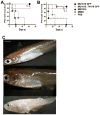
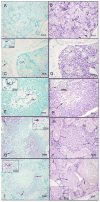
Mycobacterium ulcerans causes Buruli ulcer in humans, a progressive ulcerative epidermal lesion due to the mycolactone toxin produced by the bacterium. Molecular analysis of M. ulcerans reveals it is closely related to Mycobacterium marinum, a pathogen of both fish and man. Molecular evidence from diagnostic PCR assays for the insertion sequence IS2404 suggests an association of M. ulcerans with fish. However, fish infections by M. ulcerans have not been well documented and IS2404 has been found in other mycobacteria. We have thus, employed two experimental approaches to test for M. ulcerans in fish. We show here for the first time that M. ulcerans with or without the toxin does not mount acute or chronic infections in Japanese Medaka "Oryzias latipes" even at high doses. Moreover, M. ulcerans-infected medaka do not exhibit any visible signs of infection nor disease and the bacteria do not appear to replicate over time. In contrast, similar high doses of the wild-type M. marinum or a mycolactone-producing M. marinum "DL" strain are able to mount an acute disease with mortality in medaka. Although these results would suggest that M. ulcerans does not mount infections in fish we have evidence that CLC macrophages from goldfish are susceptible to mycolactones.
Copyright © 2012 Institut Pasteur. Published by Elsevier Masson SAS. All rights reserved.
Figures


Similar articles
-
Globally distributed mycobacterial fish pathogens produce a novel plasmid-encoded toxic macrolide, mycolactone F.Infect Immun. 2006 Nov;74(11):6037-45. doi: 10.1128/IAI.00970-06. Epub 2006 Aug 21. Infect Immun. 2006. PMID: 16923788 Free PMC article.
-
Mycobacterium marinum produces long-term chronic infections in medaka: a new animal model for studying human tuberculosis.Comp Biochem Physiol C Toxicol Pharmacol. 2007 Feb;145(1):45-54. doi: 10.1016/j.cbpc.2006.07.012. Epub 2006 Aug 12. Comp Biochem Physiol C Toxicol Pharmacol. 2007. PMID: 17015042 Free PMC article.
-
Evolution of Mycobacterium ulcerans and other mycolactone-producing mycobacteria from a common Mycobacterium marinum progenitor.J Bacteriol. 2007 Mar;189(5):2021-9. doi: 10.1128/JB.01442-06. Epub 2006 Dec 15. J Bacteriol. 2007. PMID: 17172337 Free PMC article.
-
The genome, evolution and diversity of Mycobacterium ulcerans.Infect Genet Evol. 2012 Apr;12(3):522-9. doi: 10.1016/j.meegid.2012.01.018. Epub 2012 Jan 28. Infect Genet Evol. 2012. PMID: 22306192 Review.
-
Buruli ulcer and mycolactone-producing mycobacteria.Jpn J Infect Dis. 2013;66(2):83-8. doi: 10.7883/yoken.66.83. Jpn J Infect Dis. 2013. PMID: 23514902 Review.
Cited by
-
Identification of Ser/Thr kinase and forkhead associated domains in Mycobacterium ulcerans: characterization of novel association between protein kinase Q and MupFHA.PLoS Negl Trop Dis. 2014 Nov 20;8(11):e3315. doi: 10.1371/journal.pntd.0003315. eCollection 2014 Nov. PLoS Negl Trop Dis. 2014. PMID: 25412098 Free PMC article.
-
Spatiotemporal analysis of mycolactone distribution in vivo reveals partial diffusion in the central nervous system.PLoS Negl Trop Dis. 2020 Dec 2;14(12):e0008878. doi: 10.1371/journal.pntd.0008878. eCollection 2020 Dec. PLoS Negl Trop Dis. 2020. PMID: 33264290 Free PMC article.
-
Workshop report: The medaka model for comparative assessment of human disease mechanisms.Comp Biochem Physiol C Toxicol Pharmacol. 2015 Dec;178:156-162. doi: 10.1016/j.cbpc.2015.06.003. Epub 2015 Jun 19. Comp Biochem Physiol C Toxicol Pharmacol. 2015. PMID: 26099189 Free PMC article.
-
Fish and amphibians as potential reservoirs of Mycobacterium ulcerans, the causative agent of Buruli ulcer disease.Infect Ecol Epidemiol. 2013;3. doi: 10.3402/iee.v3i0.19946. Epub 2013 Feb 22. Infect Ecol Epidemiol. 2013. PMID: 23440849 Free PMC article.
-
Behavioral interplay between mosquito and mycolactone produced by Mycobacterium ulcerans and bacterial gene expression induced by mosquito proximity.PLoS One. 2023 Aug 3;18(8):e0289768. doi: 10.1371/journal.pone.0289768. eCollection 2023. PLoS One. 2023. PMID: 37535670 Free PMC article.
References
-
- World Health Organization. Buruli ulcer: diagnosis of Mycobacterium ulcerans disease. World Health Organization; Geneva, Switzerland: 2004.
-
- Pettit JH, Marchette NJ, Rees RJ. Mycobacterium ulcerans infection. Clinical and bacteriological study of the first cases recognized in South East Asia. Br J Dermatol. 1966;78:187–97. - PubMed
-
- Smith JH. Epidemiologic observations on cases of Buruli ulcer seen in a hospital in the Lower Congo. Am J Trop Med Hyg. 1970;19:657–63. - PubMed
-
- Mitchell PJ, Jerrett IV, Slee KJ. Skin ulcers caused by Mycobacterium ulcerans in koalas near Bairnsdale, Australia. Pathology. 1984;16:256–60. - PubMed
Publication types
MeSH terms
Substances
Grants and funding
LinkOut - more resources
Full Text Sources
Other Literature Sources
Medical

