

Plasma and brain fatty acid profiles in mild cognitive impairment and Alzheimer's disease
- PMID: 22466064
- PMCID: PMC3409580
- DOI: 10.3233/JAD-2012-110629
Plasma and brain fatty acid profiles in mild cognitive impairment and Alzheimer's disease
Abstract
Alzheimer's disease (AD) is generally associated with lower omega-3 fatty acid intake from fish but despite numerous studies, it is still unclear whether there are differences in omega-3 fatty acids in plasma or brain. In matched plasma and brain samples provided by the Memory and Aging Project, fatty acid profiles were quantified in several plasma lipid classes and in three brain cortical regions. Fatty acid data were expressed as % composition and as concentrations (mg/dL for plasma or mg/g for brain). Differences in plasma fatty acid profiles between AD, mild cognitive impairment (MCI), and those with no cognitive impairment (NCI) were most apparent in the plasma free fatty acids (lower oleic acid isomers and omega-6 fatty acids in AD) and phospholipids (lower omega-3 fatty acids in AD). In brain, % DHA was lower only in phosphatidylserine of mid-frontal cortex and superior temporal cortex in AD compared to NCI (-14% and -12%, respectively; both p < 0.05). The only significant correlation between plasma and brain fatty acids was between % DHA in plasma total lipids and % DHA in phosphatidylethanolamine of the angular gyrus, but only in the NCI group (+0.77, p < 0.05). We conclude that AD is associated with altered plasma status of both DHA and other fatty acids unrelated to DHA, and that the lipid class-dependent nature of these differences reflects a combination of differences in intake and metabolism.
Figures
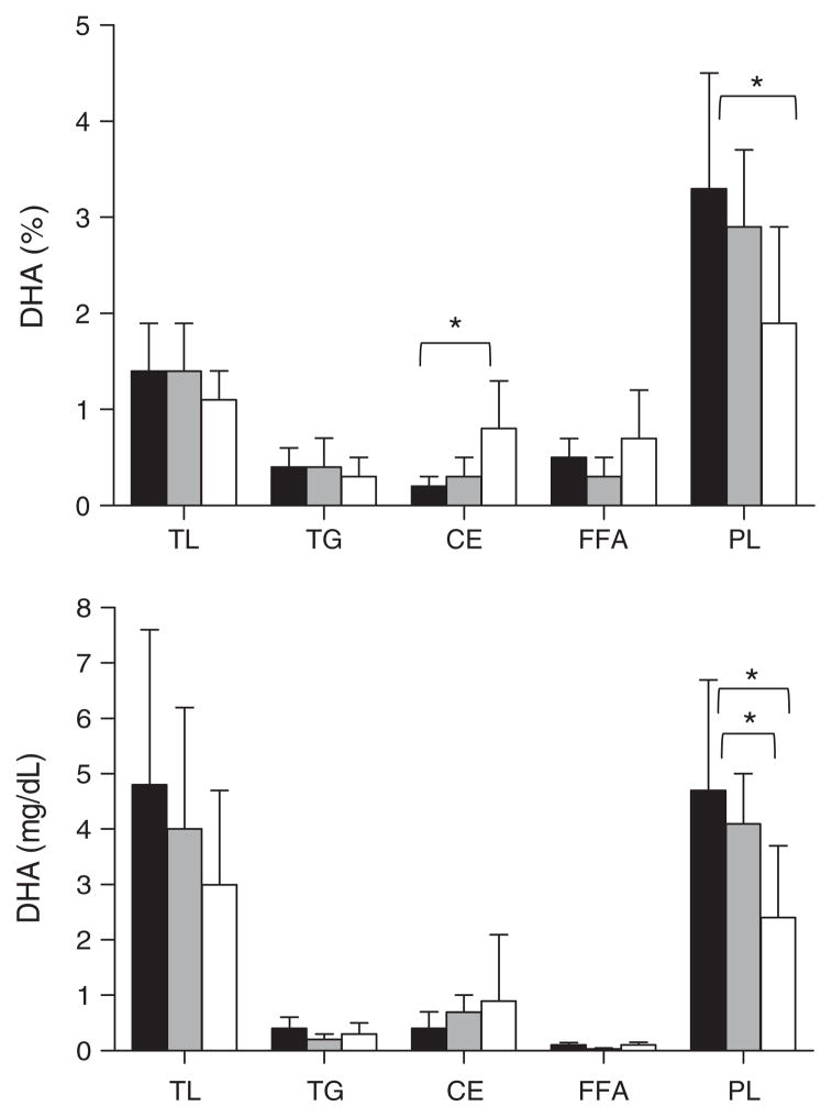
 , n = 9). Data for Alzheimer’s disease were significantly different from the no cognitive impairment group and/or the mild cognitive impairment group (*p < 0.05; Kruskal-Wallis followed by Mann-Whitney tests).
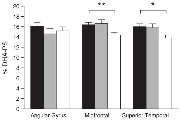 , n = 12), or Alzheimer’s disease (□, n = 12). Data for Alzheimer’s disease were significantly different from both no cognitive impairment and mild cognitive impairment groups in the midfrontal cortex (**p = 0.014; Kruskal-Wallis followed by Mann Whitney tests) and in the superior temporal cortex (*p = 0.03; Kruskal-Wallis test). No other differences in fatty acid composition for these brain regions were observed across the three groups, or across the four brain phospholipid classes studied, regardless of whether the data were expressed as concentration (mg/g) or % composition.
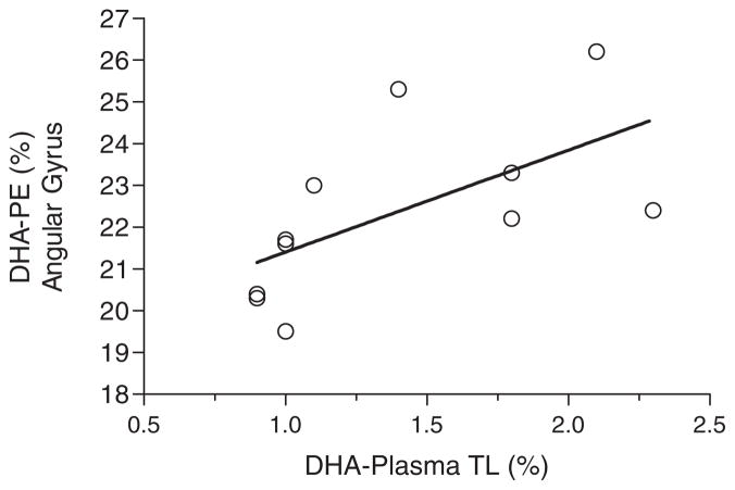
References
-
- Morris MC, Evans DA, Bienias JL, Tangney CC, Bennett DA, Wilson RS, Aggarwal N, Schneider J. Consumption of fish and n-3 fatty acids and risk of incident Alzheimer disease. Arch Neurol. 2003;60:940–946. - PubMed
-
- Gillette Guyonnet S, Abellan Van Kan G, Andrieu S, Bar-berger Gateau P, Berr C, Bonnefoy M, Dartigues JF, de Groot L, Ferry M, Galan P, Hercberg S, Jeandel C, Morris MC, Nourhashemi F, Payette H, Poulain JP, Portet F, Roussel AM, Ritz P, Rolland Y, Vellas B. IANA task force on nutrition and cognitive decline with aging. J Nutr Health Aging. 2007;11:132–152. - PubMed
-
- Boudrault C, Bazinet RP, Ma DW. Experimental models and mechanisms underlying the protective effects of n-3 polyunsaturated fatty acids in Alzheimer’s disease. J Nutr Biochem. 2009;20:1–10. - PubMed
-
- Fotuhi M, Mohassel P, Yaffe K. Fish consumption, long-chain omega-3 fatty acids and risk of cognitive decline or Alzheimer disease: A complex association. Nat Clin Pract Neurol. 2009;5:140–152. - PubMed
Publication types
MeSH terms
Substances
Grants and funding
LinkOut - more resources
Full Text Sources
Other Literature Sources
Medical
