Review
. 2012 Apr;122(4):1149-55.
doi: 10.1172/JCI58705. Epub 2012 Apr 2.

Signaling at neuro/immune synapses

Affiliations
Review

Signaling at neuro/immune synapses

Michael L Dustin. J Clin Invest. 2012 Apr.

Abstract

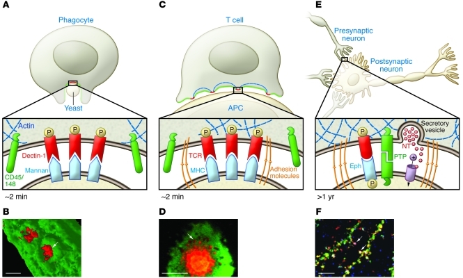
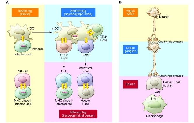
Immunological and neural synapses share properties such as the synaptic cleft, adhesion molecules, stability, and polarity. However, the mismatch in scale has limited the utility of these comparisons. The discovery of phosphatase micro-exclusion from signaling elements in immunological synapses and innate phagocytic synapses define a common functional unit at a common sub-micron scale across synapse types. Bundling of information from multiple antigen receptor microclusters by an immunological synapse has parallels to bundling of multiple synaptic inputs into a single axonal output by neurons, allowing integration and coincidence detection. Bonafide neuroimmune synapses control the inflammatory reflex. A better understanding of the shared mechanisms between immunological and neural synapses could aid in the development of new therapeutic modalities for immunological, neurological, and neuroimmunological disorders alike.


Figures

Figure 1
Figure 1. Role of submicron receptor complexes in immunological and neural synapses.
(A) Phagocytosis is triggered when CD45 and CD148 are excluded from a region more than 0.5 μm in diameter in which Syk is phosphorylated. (B) Micrograph of a phagocytic synapse. Scale bar: 4 μm. Arrow indicates the exclusion zone. Reproduced with permission from Nature (7). (C) T cell synapses are larger interfaces in which TCR microclusters that exclude CD45 are formed. These are linked by mysosin II–based contractility to augment signaling and trigger effector functions. Linkage of microclusters through myosin II ensures that T cells respond to multiple coincident MHC-peptide signals. Inset: TCR/MHC-peptide interactions with the support of adhesion molecules form microclusters that exclude CD45 and trigger robust tyrosine phosphorylation. TCR microclusters are short lived. (D) Micrograph of a T cell synapse. Scale bar: 4 μm. Arrow points to an example of a microcluster. Reproduced with permission from Nature (10). (E) Neural synapses are stabilized by adhesion molecules and can recruit receptor tyrosine kinases. More restrained signaling may promote a longer-lived junction than can then be used to process action potentials into chemical synapse and compute one output from many inputs. Eph, Eph family tyrosine kinase; PTP, protein tyrosine phosphatase; NT, neurotransmitter. (F) Microgaph of a neural synapse. Green indicates receptor-type protein tyrosine phosphatase ρ, red indicates neuroligin, blue indicates PSD-95. Scale bar: 4 μm. Arrows indicate examples of RPTPρ colocalization with neuroligin. Reproduced with permission from EMBO Journal (43).
Figure 2
Figure 2. Immunological relay race.
(A) The immune response is based on a series of immunological synapses with a common mechanism based on phosphatase exclusion. Innate leg: An intracellular pathogen infects cells, activating innate sensing mechanisms and leading to phagocytosis by an immature DC (iDC). This phagocytic synapse contributes to maturation of the DC (mDC). If the pathogen downregulates MHC class I in the infected cell, then the infected cell can be directly recognized by NK cells. Afferent leg: The mDC presents antigens on MHC class I to cytotoxic T cell precursors (CD8), on MHC class II to helper T cell precursors (CD4), and as intact complexes to B cells. Efferent leg: CTLs can directly kill MHC class I–positive infected cells, and the infected target induces cytokine production by the CD8 T cell. Helper T cells allow selection of high-affinity activated B cells and help B cells to generate an appropriate type of antibody. The B cell provides costimulatory molecules that promote cytokine production by the helper T cell. (B) The inflammatory reflex is based on innervation of a subset of helper T cells that express choline acetyltransferase. The vagus nerve relays signals to adrenergic neurons in the celiac ganglion that form neuroimmune synapses with the helper T cells. Adrenergic receptors on the T cell trigger production of acetylcholine (ACh), which interacts with cholinergic receptors on macrophages to suppress production of inflammatory cytokines such as TNF.

References

    1. Dustin ML, Colman DR. Neural and immunological synaptic relations. Science. 2002;298(5594):785–789. doi: 10.1126/science.1076386. - DOI - PubMed
    1. Norcross MA. A synaptic basis for T-lymphocyte activation. Ann Immunol (Paris). 1984;135D(2):113–134. - PMC - PubMed
    1. Paul WE, Seder RA. Lymphocyte responses and cytokines. Cell. 1994;76(2):241–251. doi: 10.1016/0092-8674(94)90332-8. - DOI - PubMed
    1. Grakoui A, et al. The immunological synapse: A molecular machine controlling T cell activation. Science. 1999;285(5425):221–227. doi: 10.1126/science.285.5425.221. - DOI - PubMed
    1. Davis DM, Chiu I, Fassett M, Cohen GB, Mandelboim O, Strominger JL. The human natural killer cell immune synapse. Proc Natl Acad Sci U S A. 1999;96(26):15062–15067. doi: 10.1073/pnas.96.26.15062. - DOI - PMC - PubMed

Publication types

MeSH terms

Substances