Review
. 2012 Apr;122(4):1164-71.
doi: 10.1172/JCI58644. Epub 2012 Apr 2.

Innate immunity in the central nervous system

Affiliations
Review

Innate immunity in the central nervous system

Richard M Ransohoff et al. J Clin Invest. 2012 Apr.

Abstract

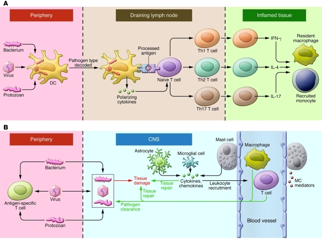
Immune responses in the CNS are common, despite its perception as a site of immune privilege. These responses can be mediated by resident microglia and astrocytes, which are innate immune cells without direct counterparts in the periphery. Furthermore, CNS immune reactions often take place in virtual isolation from the innate/adaptive immune interplay that characterizes peripheral immunity. However, microglia and astrocytes also engage in significant cross-talk with CNS-infiltrating T cells and other components of the innate immune system. Here we review the cellular and molecular basis of innate immunity in the CNS and discuss what is known about how outcomes of these interactions can lead to resolution of infection, neurodegeneration, or neural repair depending on the context.


Figures

Figure 1
Figure 1. Innate immunity in the periphery and CNS.
(A) In the face of a peripheral infection, innate immune cells prime and instruct T lymphocytes. Tissue DCs internalize microbial protein antigens, process them into peptides, and display them on their surfaces with MHC class II molecules; migrate to draining lymph nodes; and present antigens to naive CD4+ T cells. DCs direct the quality of the subsequent inflammatory response by decoding distinctive pathogen-associated signals and transmitting this information to T cells in the form of regulatory cytokines such as IL-12 (for Th1), IL-4 (Th2), or IL-6/TGF-β (Th17). In addition, lymph node environmental cues can provide information about the site of infection (gut, skin, or other). Armed with this information, effector T cells migrate to infected tissues. Upon reactivation, Th1, Th2, and Th17 cells express phenotype-defining cytokines that act on resident and recruited innate cells, which operate collectively with factors such as complement to clear the infection. (B) Resident microglia and astrocytes exert multiple functions in the CNS, including protective and restorative responses to CNS infection or injury. Cytokines and chemokines expressed by resident CNS cells also promote the recruitment of circulating lymphocytes and myeloid cells from the periphery to assist in pathogen clearance. Innate responses in the CNS cannot directly initiate adaptive immunity. Innate CNS reactions also occur during neuroinflammatory disorders and utilize many of the same components as do host defense responses.
Figure 2
Figure 2. Innate recognition of infection or tissue injury.
Endogenous DAMPs such as HSP or exogenous bacterial PAMPs activate the innate immune cells of the CNS by engaging cell surface receptors such as TLRs. Ligation of TLRs initiates an intracellular signaling cascade that involves activation of NF-κB and MAPK activity, which leads to formation of transcription factor AP1. Together these components drive transcription of IL-1 family cytokine precursor proteins pro–IL-1β and pro–IL-18. NLRs are activated by cytosolic microbial products, changes in cytosolic pH, or potassium levels often associated with stimulation of the ligand-gated ion channel P2X7R by extracellular ATP. NLRs such as NALP3 are central constituents of inflammasomes, multiprotein complexes that mediate activation of caspase-1, which catalyzes cleavage and maturation of IL-1 family cytokines. Functional domains of inflammasome components include caspase activation and recruitment domains (CARDs), which are present in both caspase-1 and the adapter apoptosis-associated Speck-like protein containing CARD (ASC). PYD, pyrin domain.
Figure 3
Figure 3. Microglial cells, as described by Pio del Rio-Hortega (161).
(A) Pio del Rio-Hortega (1882–1945), who characterized and named microglial cells. (B) Images of ramified microglial cells drawn by Hortega. (C) Morphological transformation of microglia to phagocytic macrophage. Panels as lettered in C: A, Microglial cell with modestly thickened processes as compared with ramified microglia; B, microglial cell with short, thick processes and enlarged cell body; C, microglial cell with pseudopodia; D, amoeboid microglial cell; E, amoeboid microglial cell with pseudopodia; F, microglial cell with phagocytosed leukocyte; G, microglial cell with numerous phagocytosed erythrocytes; H, microglial cell with lipid inclusions, also termed “foam cell”; I, microglial cell in mitotic division. Reproduced with permission from Physiological Reviews (161).

References

    1. Edele F, et al. Cutting edge: instructive role of peripheral tissue cells in the imprinting of T cell homing receptor patterns. J Immunol. 2008;181(6):3745–3749. - PubMed
    1. Dudda JC, et al. Dendritic cells govern induction and reprogramming of polarized tissue-selective homing receptor patterns of T cells: important roles for soluble factors and tissue microenvironments. Eur J Immunol. 2005;35(4):1056–1065. doi: 10.1002/eji.200425817. - DOI - PubMed
    1. Mora JR, Cheng G, Picarella D, Briskin M, Buchanan N, von Andrian UH. Reciprocal and dynamic control of CD8 T cell homing by dendritic cells from skin- and gut-associated lymphoid tissues. . J Exp Med. 2005;201(2):303–316. doi: 10.1084/jem.20041645. - DOI - PMC - PubMed
    1. Oyoshi MK, et al. Epicutaneous challenge of orally immunized mice redirects antigen-specific gut-homing T cells to the skin. J Clin Invest. 2011;121(6):2210–2220. - PMC - PubMed
    1. Galea I, Bechmann I, Perry VH. What is immune privilege (not)? Trends Immunol. 2007;28(1):12–18. doi: 10.1016/j.it.2006.11.004. - DOI - PubMed

Publication types

MeSH terms