. 2012 Aug;27(8):1695-707.
doi: 10.1002/jbmr.1623.

V-ATPase subunit ATP6AP1 (Ac45) regulates osteoclast differentiation, extracellular acidification, lysosomal trafficking, and protease exocytosis in osteoclast-mediated bone resorption

Affiliations

V-ATPase subunit ATP6AP1 (Ac45) regulates osteoclast differentiation, extracellular acidification, lysosomal trafficking, and protease exocytosis in osteoclast-mediated bone resorption

De-Qin Yang et al. J Bone Miner Res. 2012 Aug.

Abstract

Lysosomal trafficking and protease exocytosis in osteoclasts are essential for ruffled border formation and bone resorption. Yet the mechanism underlying lysosomal trafficking and the related process of exocytosis remains largely unknown. We found ATP6ap1 (Ac45), an accessory subunit of vacuolar-type H(+)-ATPases (V-ATPases), to be highly induced by receptor activator for nuclear factor kappa B ligand (RANKL) in osteoclast differentiation. Ac45 knockdown osteoclasts formed normal actin rings, but had severely impaired extracellular acidification and bone resorption. Ac45 knockdown significantly reduced osteoclast formation. The decrease in the number of osteoclasts does not result from abnormal apoptosis; rather, it results from decreased osteoclast precursor cell proliferation and fusion, which may be partially due to the downregulation of extracellular signal-regulated kinase (ERK) phosphorylation and FBJ osteosarcoma oncogene (c-fos), nuclear factor of activated T-cells, cytoplasmic 1 (NFATc1), and "transmembrane 7 superfamily member 4" (Tm7sf4) expression. Notably, Ac45 knockdown osteoclasts exhibited impaired lysosomal trafficking and exocytosis, as indicated by the absence of lysosomal trafficking to the ruffled border and a lack of cathepsin K exocytosis into the resorption lacuna. Our data revealed that the impaired exocytosis is specifically due to Ac45 deficiency, and not the general consequence of a defective V-ATPase. Together, our results demonstrate the essential role of Ac45 in osteoclast-mediated extracellular acidification and protease exocytosis, as well as the ability of Ac45 to guide lysosomal intracellular trafficking to the ruffled border, potentially through its interaction with the small guanosine-5'-triphosphatase (GTPase) Rab7. Our work indicates that Ac45 may be a novel therapeutic target for osteolytic disease.


Conflict of interest statement

Disclosures

All authors state that they have no conflicts of interest.

Figures

Fig. 1
Fig. 1
Expression of Ac45 in osteoclasts and its effective depletion by lentiviral siRNA. (A) Microarray data of expression levels of Ac45 in monocytes and osteoclasts. Intensity units (IU). (B) Western blot analysis of the time-course of Ac45 protein expression in MBM induced by M-CSF and RANKL. (C) The protein levels on time-course blot were analyzed and quantified with the NIH ImageJ software. The values shown represent ratios of Ac45 to β-actin protein levels that have been normalized (n=3). (D) Western blot verified Ac45 knockdown effect of 5 siRNAs by lentivirus-mediated transduction. GAPDH was used as a protein loading control and Lentivirus-GFP-RNAi as a transduction efficiency control. (E) The protein levels on blot were analyzed and quantified with the NIH ImageJ software. The values shown represent ratios of Ac45 to GAPDH protein levels that have been normalized (n=3). *P<0.05, **P<0.01 compared with that of Lentivirus-GFP-RNAi treated cells.
Fig. 2
Fig. 2
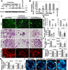
Lentivirus-mediated knockdown of Ac45 reduced the formation of multinucleated osteoclasts, but it did not impair actin rings formation. (A) Western blot of Ac45 protein expression in mock cells and monocytes, as well as in osteoclasts transduced with Lentivirus-Ac45-RNAi (s1) and Lentivirus-GFP-RNAi after 48 or 72 hours of M-CSF/RANKL induction. β-actin was used as a protein loading control. (B) The protein levels on blot were analyzed and quantified with the NIH ImageJ software. The values shown represent ratios of Ac45 to β-actin protein levels that have been normalized (n=3). (C) Various assays performed on osteoclasts transduced with Lentivirus-GFP-RNAi, Lentivirus-Ac45-RNAi(s1), or Lentivirus-Ac45-RNAi(s2). Row 1: Verification of lentivirus transduction and siRNA expression through GFP reporter gene expression. Row 2 and 3: TRAP staining of osteoclasts transduced after 48 or 72 hours of M-CSF/RANKL induction as indicated. Row 4: Immunostaining of F-actin podosomal belts with rhodamine phalloidin. The osteoclasts shown were representative of the data (n=3). (D) Quantification of TRAP-positive multinucleated cell (MNC) number (≥ 3 nuclei) in rows 2 & 3 of C. All data are expressed as mean ± s.d. (n=5). (E) Quantification of the number of nuclei per MNC in rows 2 & 3 of C. All data are expressed as mean ± s.d. (n=10). (F) Quantification of MNC size in rows 2 & 3 of C. All data are expressed as mean ± s.d. (n=10). (G) d2 mRNA expression level relative to Hprt1. (n=3). (H) Tm7sf4 mRNA expression level relative to Hprt1. (n=3). In D-H, column 1 is Lentivirus-GFP-RNAi, column 2 is Lentivirus-Ac45-RNAi(s1), and column 3 is Lentivirus-Ac45-RNAi(s2). (I) Apoptosis assay with Hoechst 33258 staining in osteoclasts 48 hours after M-CSF/RANKL induction. Compared to the positive apoptosis control (apoptosis induced by M-CSF/RANKL starvation for 12 hours), apoptosis does not occur as a result of Ac45 depletion. Red arrows indicate apoptotic nuclei and white arrows indicate normal nuclei. *P<0.05, **P<0.01, ***P<0.001 compared with that of Lenti-GFPi treated cells at the same time points of transduction.
Fig. 3
Fig. 3
Depletion of Ac45 impaired extracellular acidification, V-ATPase trafficking to the plasma membrane, and osteoclastic bone resorption. (A) Acridine orange staining of mock osteoclasts and osteoclasts transduced by Lentivirus-GFP-RNAi, Lentivirus-Ac45-RNAi(s1), Lentivirus-Ac45-RNAi(s2), and Lentivirus-ATP6v1e1-RNAi (as a control) after 48 or 72 hours of M-CSF/RANKL induction. (B) Atp6v1c1 and Atp6v0a3 immunofluorescence staining of osteoclasts as indicated, white arrows indicate colocalization of Atp6v1c1 and Atp6v0a3. (C) TRAP staining of bone discs with osteoclasts as indicated. (D) Rhodamine phalloidin and DAPI staining of osteoclasts on bone as indicated. (E) Quantification of nuclei per mm2 in D (n=5). (F) Quantification of MNCs’ nuclei per mm2 in D (n=5). (G) ELISA assay of CTX concentration in osteoclast culture media as indicated. (H) Bone resorption pits on bovine cortical bone slices resulting from mock osteoclasts and osteoclasts transduced with Lentivirus-GFP-RNAi, Lentivirus-Ac45-RNAi(s1), and Lentivirus-Ac45-RNAi(s2) after 48 or 72 hours of M-CSF/RANKL induction. The bone slices were subjected to scanning electron microscopy analysis. White inset boxes: higher magnification view of the bone resorption pits. (I) Quantification of percentage of resorption area per mm2 among the groups at different time points of transduction. (n=3). In E, F, G, and I, column 1 is Mock, column 2 is Lentivirus-GFP-RNAi, column 3 is Lentivirus-Ac45-RNAi(s1), and column 4 is Lentivirus-Ac45-RNAi(s2). *P<0.05, **P<0.01 compared with that of Lentivirus-GFP-RNAi treated cells. All results are mean ± s.d.
Fig. 4
Fig. 4
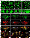
Depletion of Ac45 resulted in sorting failure and a loss of lysosomal trafficking to the ruffled border. (I) Three different antibodies anti-ATP6v0a3, anti-ATP6v1b2, and anti-ATP6v1c1 were used to localize V-ATPases either in (IA-Ii) non-resorptive cells on glass slices or (IJ-IU) resorptive cells on dentin slices. Mock osteoclasts and osteoclasts transduced with Lentivirus-GFP-RNAi or Lentivirus-Ac45-RNAi(s1) after induction with M-CSF/RANKL for 48 hours were viewed as follows: (IA-Ii) Zeiss Axioplan microscope assay of V-ATPase localization; (IJ-IR) horizontal views (x-y sections) of confocal microscopy analysis of V-ATPase localization; (IJz-IRz) lateral views (z-x sections) of cells stained for anti-ATP6v0a3, anti-ATP6v1b2, and anti-ATP6v1c1. The positions of confocal sections are shown by dotted lines in the corresponding images. (IS-IU) Three-dimensional reconstruction of the cell on the bone surface. The length of the scale bar is 10 µm. (II) Two different antibodies, anti-ATP6v0a3 (fluorescence appears green) and anti-lamp-1 (fluorescence appears red), were used to localize lysosomes in resorptive cells [mock osteoclasts and osteoclasts transduced with Lentivirus-GFP-RNAi or Lentivirus-Ac45-RNAi(s1)] on bone slices. (IIA-IIi) Three-dimensional reconstruction of cells on the bone surface. Nuclei were labeled using DAPI DNA stain, and appear blue in merged images. Yellow staining in merged images indicates colocalization of Atp6v0a3 and lamp-1. (IIJ-IIL) Slices and sections of IIG-IIi. The surface of the cells in XZ and YZ is outlined in white. The positions of confocal sections are shown by dotted lines in the corresponding images. White arrows indicate staining at the ruffled border. Blue arrows indicate staining in monocytes. The length of the scale bar is 20 µm.
Fig. 5
Fig. 5
Depletion of Ac45 causes defects in cathepsin K exocytosis. Cathepsin K was immunostained with anti-cathepsin K antibody and then costained with F-actin by secondary antibody conjugated with FITC and rhodamine phalloidin in (A-C) mock cells and osteoclasts transduced with (D-F, Dz-Fz) Lentivirus-GFP-RNAi, (G-I) Lentivirus-Ac45-RNAi(s1), (J-L, Jz-Lz) Lentivirus-Ac45-RNAi(s2), or (M-O) Lentivirus-ATP6v1e1-RNAi (as a control) after induction with M-CSF/RANKL for 48 hours. These resorbing cells were detected by confocal microscopy and analyzed by three-dimensional reconstruction software Imaris. Horizontal views (x-y sections) of 1-µm thickness were taken 1-µm above the bone surface. Lateral views (z-x sections) of (Dz-Fz) mock cells and (Jz-Lz) osteoclasts transduced with Lentivirus-Ac45-RNAi(s2) that were stained for the cathepsin K and actin. The lateral view reveals that unlike mock cells, osteoclasts transduced with Lentivirus-Ac45-RNAi(s2) do not form deep bone resorption pits as indicated by the white arrows and lines. The positions of confocal sections are shown by dotted lines in the corresponding images. The length of the scale bar is 10 µm. (P) Representative Western blotting assay of cathepsin K protein level in osteoclast culture media as indicated. (Q) Representative ponceau S stain for Western blots of P. (R) Quantification of P. (n=3). In P-R, column 1 is Mock, column 2 is Lentivirus-GFP-RNAi, column 3 is Lentivirus-Ac45-RNAi(s1), column 4 is Lentivirus-Ac45-RNAi(s2). M indicates marker. **P<0.01 compared with that of Lentivirus-GFP-RNAi treated cells. All results are mean ± s.d.
Fig. 6
Fig. 6
Ac45 interacts with Rab7 in osteoclasts. (A) Ac45 colocalized with Rab7 (white arrows indicate colocalization) in resorbing osteoclasts cultured on bone slice. The cells shown are representative of the data. (B) Immunoblot of Ac45 when the whole cell extracts were immunoprecipitated with anti-Rab7 and anti-Rac1. (C) Top Left: Immunoprecipitation with anti-FLAG of cells transduced with recombinant viruses containing an empty vector as a control (vec), wild-type Rab7 (Rab7WT) and the active form of Rab7 (Rab7Q67L). The cells were analyzed by Western blot for the detection of Ac45 and IgG (positive control). Top Right: Total cell lysates (TCL) of non-immunoprecipitated cells were analyzed with Western blot for Ac45 and IgG (negative control). Bottom: Immunoprecipitated cells and total cell lysates analyzed by Western blot for FLAG (control).
Fig. 7
Fig. 7
Lentivirus-mediated knockdown of Ac45 impaired differentiation of mononuclear cells to mature osteoclasts, inhibited osteoclast precursor cell proliferation, and downregulated proteins that promote osteoclastogensis, but had no effect on osteoclast precursor cell apoptosis. (A) TRAP staining of mock cells, as well as osteoclasts transduced with Lentivirus-GFP-RNAi, Lentivirus-Ac45-RNAi(s1), and Lentivirus-Ac45-RNAi(s2) after 12 or 24 hours of M-CSF/RANKL induction. (B) Quantification of TRAP+ multinucleated cell (MNC) number (≥ 3 nuclei) in A. All data are expressed as mean ± s.d. (n=5). (C)Anti-BrdU staining of osteoclast precursor cells after 2.5 hours of incubation with BrdU and M-CSF. (D) Quantification of the percentage of BrdU positive cells per view. (n=10). (E) TUNEL staining of osteoclast precursor cells after 24 hours of incubation with media without FBS and M-CSF. (F) Quantification of the ratio of TUNEL positive cells per view. (n=10). In B, D, and F, column 1 is Mock, column 2 is Lentivirus-GFP-RNAi, column 3 is Lentivirus-Ac45-RNAi(s1), and column 4 is Lentivirus-Ac45-RNAi(s2). **P<0.01, ***P<0.001 compared with that of Lentivirus-GFP-RNAi treated cells at the same time points of transduction. (G) Western blot analysis of expression levels of ERK, phosphorylated ERK (p-ERK), c-fos, and NFATc1 in mock osteoclasts and osteoclasts transduced by Lentivirus-GFP-RNAi (control), Lentivirus-Ac45-RNAi(s1), or Lentivirus-Ac45-RNAi(s2) after 24 or 72 hours of M-CSF/RANKL induction. β-actin was used as a protein loading control. (H) Quantification of the protein levels on blot (lanes 1–7). The values shown represent ratios of ERK, p-ERK, c-fos, or NFATc1 to β-actin protein levels that have been normalized (n=3).

Similar articles

Cited by

References

    1. Rousselle AV, Heymann D. Osteoclastic acidification pathways during bone resorption. Bone. 2002 Apr;30(4):533–540. - PubMed
    1. Akisaka T, Yoshida H, Suzuki R, Takama K. Adhesion structures and their cytoskeleton-membrane interactions at podosomes of osteoclasts in culture. Cell Tissue Res. 2008 Mar;331(3):625–641. - PubMed
    1. Palokangas H, Mulari M, Vaananen HK. Endocytic pathway from the basal plasma membrane to the ruffled border membrane in bone-resorbing osteoclasts. J.Cell Sci. 1997 Aug;110(Pt 15):1767–1780. - PubMed
    1. Mulari MT, Zhao H, Lakkakorpi PT, Vaananen HK. Osteoclast ruffled border has distinct subdomains for secretion and degraded matrix uptake. Traffic. 2003 Feb;4(2):113–125. - PubMed
    1. Mellman I, Nelson WJ. Coordinated protein sorting, targeting and distribution in polarized cells. Nat.Rev.Mol Cell Biol. 2008 Nov;9(11):833–845. - PMC - PubMed

Publication types

MeSH terms