IL1β induces mesenchymal stem cells migration and leucocyte chemotaxis through NF-κB
- PMID: 22467443
- PMCID: PMC3412085
- DOI: 10.1007/s12015-012-9364-9
IL1β induces mesenchymal stem cells migration and leucocyte chemotaxis through NF-κB
Abstract
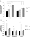
Mesenchymal stem cells are often transplanted into inflammatory environments where they are able to survive and modulate host immune responses through a poorly understood mechanism. In this paper we analyzed the responses of MSC to IL-1β: a representative inflammatory mediator. Microarray analysis of MSC treated with IL-1β revealed that this cytokine activateds a set of genes related to biological processes such as cell survival, cell migration, cell adhesion, chemokine production, induction of angiogenesis and modulation of the immune response. Further more detailed analysis by real-time PCR and functional assays revealed that IL-1β mainly increaseds the production of chemokines such as CCL5, CCL20, CXCL1, CXCL3, CXCL5, CXCL6, CXCL10, CXCL11 and CX(3)CL1, interleukins IL-6, IL-8, IL23A, IL32, Toll-like receptors TLR2, TLR4, CLDN1, metalloproteins MMP1 and MMP3, growth factors CSF2 and TNF-α, together with adhesion molecules ICAM1 and ICAM4. Functional analysis of MSC proliferation, migration and adhesion to extracellular matrix components revealed that IL-1β did not affect proliferation but also served to induce the secretion of trophic factors and adhesion to ECM components such as collagen and laminin. IL-1β treatment enhanced the ability of MSC to recruit monocytes and granulocytes in vitro. Blockade of NF-κβ transcription factor activation with IκB kinase beta (IKKβ) shRNA impaired MSC migration, adhesion and leucocyte recruitment, induced by IL-1β demonstrating that NF-κB pathway is an important downstream regulator of these responses. These findings are relevant to understanding the biological responses of MSC to inflammatory environments.
Figures


References
-
- Pereira RF, O'Hara MD, Laptev AV, Halford KW, Pollard MD, Class R, Simon D, Livezey K, Prockop DJ. Marrow stromal cells as a source of progenitor cells for nonhematopoietic tissues in transgenic mice with a phenotype of osteogenesis imperfecta. Proceedings of the National Academy of Sciences of the United States of America. 1998;95:1142–7. doi: 10.1073/pnas.95.3.1142. - DOI - PMC - PubMed
-
- Gnecchi M, He H, Noiseux N, Liang OD, Zhang L, Morello F, Mu H, Melo LG, Pratt RE, Ingwall JS, Dzau VJ. Evidence supporting paracrine hypothesis for Akt-modified mesenchymal stem cell-mediated cardiac protection and functional improvement. The FASEB Journal. 2006;20:661–9. doi: 10.1096/fj.05-5211com. - DOI - PubMed
Publication types
MeSH terms
Substances
LinkOut - more resources
Full Text Sources
Other Literature Sources
Molecular Biology Databases
Research Materials
Miscellaneous

