. 2012 Jun;194(11):2949-61.
doi: 10.1128/JB.06820-11. Epub 2012 Mar 30.

The PhoP/PhoQ system and its role in Serratia marcescens pathogenesis

Affiliations

The PhoP/PhoQ system and its role in Serratia marcescens pathogenesis

Julieta Barchiesi et al. J Bacteriol. 2012 Jun.

Abstract

Serratia marcescens is able to invade, persist, and multiply inside nonphagocytic cells, residing in nonacidic, nondegradative, autophagosome-like vacuoles. In this work, we have examined the physiological role of the PhoP/PhoQ system and its function in the control of critical virulence phenotypes in S. marcescens. We have demonstrated the involvement of the PhoP/PhoQ system in the adaptation of this bacterium to growth on scarce environmental Mg(2+), at acidic pH, and in the presence of polymyxin B. We have also shown that these environmental conditions constitute signals that activate the PhoP/PhoQ system. We have found that the two S. marcescens mgtE orthologs present a conserved PhoP-binding motif and demonstrated that mgtE1 expression is PhoP dependent, reinforcing the importance of PhoP control in magnesium homeostasis. Finally, we have demonstrated that phoP expression is activated intracellularly and that a phoP mutant strain is defective in survival inside epithelial cells. We have shown that the Serratia PhoP/PhoQ system is involved in prevention of the delivery to degradative/acidic compartments.


Figures

Fig 1
Fig 1
(A) PhoP binding sites in Serratia marcescens genome. A consensus motif for the PhoP binding site was generated using a collection of previously defined promoter regions of PhoP-regulated genes in S. Typhimurium and E. coli as a database and the MEME software tool (3). The logo shows the consensus motif for the PhoP binding site (weblogo.berkeley.edu), and the putative PhoP binding site sequences with high scores identified in the S. marcescens Db11 genome by MAST (4) are listed below. The PhoP boxes are shown in boldface; arrows indicate orientation relative to each translational start site. The name of the gene and the distance from the translational start site are also indicated. (B, C, and D) Mg2+, pH, and polymyxin B control PhoP expression in S. marcescens. β-Galactosidase activity from the phoP::lacZ transcriptional fusion was determined. Bacteria were grown overnight in LB or N medium with 10 μM MgCl2 for low Mg2+ and LB plus 50 mM MgCl2 or N medium plus 1 and 10 mM MgCl2 for high Mg2+ (B) in N medium MES (pH 7.7) to exponential phase and then inoculated into N medium MES (pH 7.7 or 5.5) and grown for an additional 4 h (C) or into LB or N medium with 1 mM MgCl2 to exponential phase and then inoculated into LB and LB plus 100 μg/ml polymyxin B or N medium with 1 mM MgCl2 and N medium with 1 mM MgCl2 plus 20 μg/ml polymyxin B, grown for 1 h (D). The data correspond to mean values for four independent experiments performed in duplicate in each case. Error bars correspond to standard deviations. (∗, P < 0.001).
Fig 2
Fig 2
PhoP is required for Serratia to resist an environment deprived of Mg2+, with low pH, or with the presence of polymyxin B. (A) WT and phoP Serratia strains harboring the pJB and pJB::phoPQ plasmids were grown in N minimal medium supplemented with 10 μM or 10 mM MgCl2, and the OD600 was determined every 60 min. (B) WT and phoP Serratia cells harboring the pJB and pJB::phoPQ plasmids were grown in in E glucose broth at pH 7 or pH 5, and the OD600 was determined every 60 min. (C) The OD600 was determined for overnight cultures of the WT and phoP Serratia strains grown in LB in the presence of different concentrations of polymyxin B. (D) The OD600 was determined on overnight cultures of the SmDb11 WT and phoP strains grown in N minimal medium supplemented with 10 μM MgCl2 or LB with 6 mg/ml of polymyxin B. For the acid resistance assay, both strains were grown in E glucose broth at pH 4.3, and the OD600 was determined after 6 h. Results are averages from three independent assays performed in duplicate. Error bars correspond to standard deviations. (∗, P < 0.001).
Fig 3
Fig 3
mgtE1 responds to the same signals as phoP. (A) Schematic representation of the promoter region of mgtE1, including the predicted PhoP binding site (in black), the riboswitch-encompassed region (M-Box; the cartoon does not reflect an actual secondary structure but depicts only the localization of the predicted structure), the insertion site of mini-Tn5-lacZ, the predicted −35 and −10 regions (underlined), and the predicted transcriptional (bold G) and translational (boxed ATG) start site. (B, C, and D) β-Galactosidase activity from the mgtE1::lacZ transcriptional fusion was determined. Strains were grown overnight in LB for low Mg2+ and LB plus 50 mM MgCl2 for high Mg2+ (B) and in N medium MES (pH 7.7) to exponential phase and then inoculated into N medium MES (pH 7.7 or 5.5) and grown additional 4 h (C) or in LB to exponential phase and then inoculated into LB and LB plus 20 μg/ml polymyxin B and grown for 1 h (D). Data correspond to mean values for four independent experiments performed in duplicate; error bars correspond to standard deviations (∗, P < 0.001; ∗∗, P > 0.2).
Fig 4
Fig 4
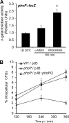
PhoP is involved in S. marcescens survival inside epithelial cells. (A) CHO cells were infected with the phoP::lacZ strain. At 180 min p.i., cells were disrupted, and β-galactosidase activity from intracellular bacteria was determined. β-Galactosidase values obtained from the phoP::lacZ strain grown in α-MEM culture medium and LB medium during the same time lapse were determined as the control. Values correspond to the ratio between the β-galactosidase activity obtained under each condition and the activity for the same strain grown in LB. Values are the means for triplicate wells from a representative of three independent experiments. Error bars correspond to standard deviations. β-Galactosidase activity values from intracellular bacteria and bacteria in α-MEM or LB were significantly different (∗, P < 0.001). (B) CHO cells were infected with the WT/pJB, phoP/pJB, and phoP/pJB::phoPQ strains. Percentages of intracellular CFU calculated relative to CFU in the inoculum were determined at the indicated times. Results are averages from at least three independent assays performed in duplicate. Error bars correspond to standard deviations.
Fig 5
Fig 5
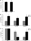
The phoP mutant is mainly delivered to an acidic and degradative autophagic compartment. (A) CHO cells were infected with the WT/pGFP and phoP/pGFP strains at an MOI of 10 for 30 min. Then, cells were fixed and extracellular Serratia was stained using anti-Serratia antibodies and detected by incubation with anti-rabbit Cy3-conjugated secondary antibodies. Bacterial internalization was calculated as the number of intracellular bacteria per cell. In each case, cells were analyzed by confocal microscopy. At least 200 infected cells (B and C) or 600 cells (A) were scored from three independent experiments. Data represent the means for three independent experiments. Error bars correspond to standard deviations (∗∗, P > 0.2). (B) CHO cells were infected with the WT/pGFP and phoP/pGFP strains. After 360 min p.i., cells were fixed and intracellular bacteria were detected by fluorescence. To label acidic compartments, after 60 min p.i., cells were incubated with LysoTracker. To assay degradative compartments, CHO cells were preincubated with DQ-BSA. In a third independent assay, the lysosomal protease cathepsin D was detected using rabbit anti-human cathepsin D and secondary antibody goat anti-rabbit IgG conjugated with Cy3. The percentage of colocalization was calculated as the ratio between the number of vesicles containing bacteria that colocalize with the indicated fluorescently labeled marker and the total number of SeCVs. Error bars correspond to standard deviations (∗, P < 0.001). (C) CHO-EGFP-LC3 cells were infected with WT or phoP strain. After 60 min p.i., cells were incubated with LysoTracker to label acidic compartments. Cells were fixed at 360 min p.i., and intracellular bacteria were detected with anti-Serratia antibodies coupled with a secondary antibody labeled with Alexa Fluor 647. The percentages of colocalization of bacteria with EGFP-LC3 and/or with LysoTracker were calculated as the ratio between the number of autophagic SeCVs that colocalize with LysoTracker (acidic) or not (nonacidic) and the total number of autophagic SeCVs. Error bars correspond to standard deviations (∗, P < 0.001).
Fig 6
Fig 6
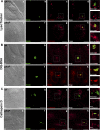
Representative confocal microscopy images of CHO cells infected with S. marcescens WT and phoP strains. (A) CHO cells infected with the WT/pGFP and phoP/pGFP strains (green fluorescence; b and f) and incubated with LysoTracker (red fluorescence, c and g) at 360 min p.i. are shown. Insets show higher magnification of the boxed area in the merged and LysoTracker images, respectively. The images highlight a nonacidic SeCV for the WT strain and an acidic SeCV for the phoP mutant strain. (B) CHO cells preincubated with DQ-BSA (red fluorescence; c and g) and infected with WT/pGFP and phoP/pGFP strains (green fluorescence; b and f) at 360 min p.i. are shown. Insets show higher magnification of the boxed area in the merged and DQ-BSA images, respectively. The images highlight a nondegradative SeCV for the WT strain and a degradative SeCV for the phoP mutant strain. (C) CHO cells infected with WT/pGFP and phoP/pGFP strains (green fluorescence; b and f) at 360 min p.i. are shown. Cathepsin D was detected with anti-cathepsin D antibodies coupled with a secondary antibody labeled with Cy3 (red fluorescence; c and g). Inset images show higher magnification of the boxed area in the merged and cathepsin D images, respectively, and highlight a SeCV without a lysosomal marker for the WT strain and a SeCV containing a lysosomal marker for the phoP mutant strain.

References

    1. Alexeyev MF. 1999. The pKNOCK series of broad-host-range mobilizable suicide vectors for gene knockout and targeted DNA insertion into the chromosome of gram-negative bacteria. Biotechniques 26:824–826, 828 - PubMed
    1. Anderson GG, Yahr TL, Lovewell RR, O'Toole GA. 2010. The Pseudomonas aeruginosa magnesium transporter MgtE inhibits transcription of the type III secretion system. Infect. Immun. 78:1239–1249 - PMC - PubMed
    1. Bailey TL, Elkan C. 1995. The value of prior knowledge in discovering motifs with MEME. Proc. Int. Conf. Intell. Syst. Mol. Biol. 3:21–29 - PubMed
    1. Bailey TL, Gribskov M. 1998. Methods and statistics for combining motif match scores. J. Comput. Biol. 5:211–221 - PubMed
    1. Bearson BL, Wilson L, Foster JW. 1998. A low pH-inducible, PhoPQ-dependent acid tolerance response protects Salmonella typhimurium against inorganic acid stress. J. Bacteriol. 180:2409–2417 - PMC - PubMed

Publication types

MeSH terms

LinkOut - more resources