. 2012 Jul 15;125(Pt 14):3454-63.
doi: 10.1242/jcs.102061. Epub 2012 Mar 30.

Polycomb repressive complex 2 impedes intestinal cell terminal differentiation

Affiliations

Polycomb repressive complex 2 impedes intestinal cell terminal differentiation

Yannick D Benoit et al. J Cell Sci. .

Abstract

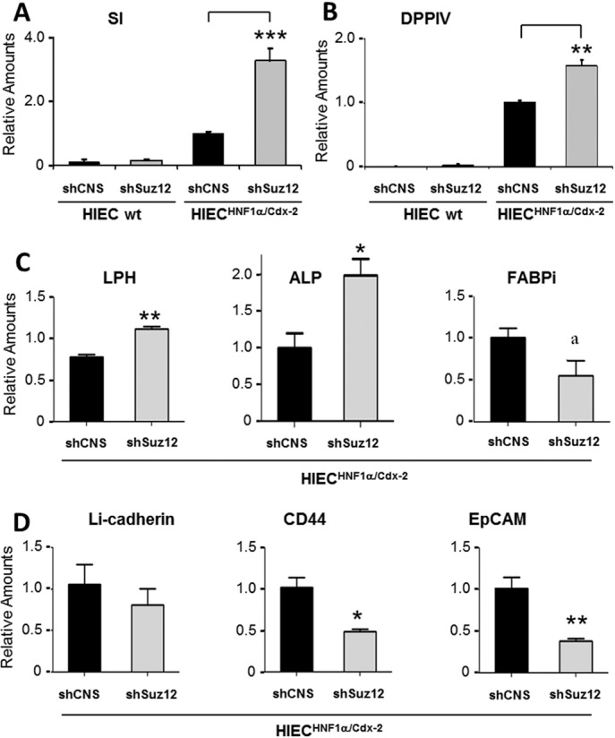
The crypt-villus axis constitutes the functional unit of the small intestine, where mature absorptive cells are confined to the villi, and stem cells and transit amplifying and differentiating cells are restricted to the crypts. The polycomb group (PcG) proteins repress differentiation and promote self-renewal in embryonic stem cells. PcGs prevent transcriptional activity by catalysing epigenetic modifications, such as the covalent addition of methyl groups on histone tails, through the action of the polycomb repressive complex 2 (PRC2). Although a role for PcGs in the preservation of stemness characteristics is now well established, recent evidence suggests that they may also be involved in the regulation of differentiation. Using intestinal epithelial cell models that recapitulate the enterocytic differentiation programme, we generated a RNAi-mediated stable knockdown of SUZ12, which constitutes a cornerstone for PRC2 assembly and functionality, in order to analyse intestinal cell proliferation and differentiation. Expression of SUZ12 was also investigated in human intestinal tissues, revealing the presence of SUZ12 in most proliferative epithelial cells of the crypt and an increase in its expression in colorectal cancers. Moreover, PRC2 disruption led to a significant precocious expression of a number of terminal differentiation markers in intestinal cell models. Taken together, our data identified a mechanism whereby PcG proteins participate in the repression of the enterocytic differentiation program, and suggest that a similar mechanism exists in situ to slow down terminal differentiation in the transit amplifying cell population.


Figures

Fig. 1.
Fig. 1.
Impact of SUZ12 knockdown on the Caco-2/15 differentiation program. (A) Western blot analysis demonstrates the expression profile of SI and SUZ12 proteins in 0-, 8-, 15- and 30-day post-confluent Caco-2/15 cultures. Relative optical density values of immunodetected SI and SUZ12 at each confluence stage in Caco-2/15 as compared to the actin loading control are shown on the right (n = 3, *P<0.001). (B) A representative western blot for the assessment of SUZ12 protein expression levels and the PRC2-associated epigenetic marker H3K27me3, in Caco-2/15 stable SUZ12 knockdown (shSUZ12) and control (shLUC) cell populations at 0, 4 and 8 days of postconfluence. Actin was used as the loading control. (C) Representative western blot of SI expression as a function of confluence (+0, +4 and +8 days) in control (shLUC) and SUZ12 knockdown (shSUZ12) Caco-2/15 cultures. (D) Amounts of SI at each confluence stage in control (shLUC) and SUZ12 knockdown (shSUZ12) Caco-2/15 cultures relative to the loading control actin (n = 3, ***P<0.0001). (E,F) Relative amounts of (E) HNF1α protein in Caco-2/15 at 0, 4 and 8 days postconfluence and (F) CDX2, E-cadherin and villin in Caco-2/15 cultures at 8 days post-confluence. Values were obtained by optical density measurements of western blot analyses, relative to the loading control actin, in control (shLUC) and SUZ12 knockdown (shSUZ12) stable SUZ12 knockdown (shSUZ12; n = 3).
Fig. 2.
Fig. 2.
SI transcript levels following SUZ12 knockdown in Caco-2 cells relative to their fully differentiated counterparts and pure intestinal epithelium. Relative amounts of SI transcript were determined by quantitative PCR in control (shLUC, black columns) and SUZ12 knockdown (shSUZ12, white columns) Caco-2/15 cells at 0, 4 and 8 days postconfluence compared to wild-type Caco-2/15 cells at subconfluence (SC), confluence (C), 3, 12 and 30 days of post confluence (curve). Amounts were determined relative to the average of five purified intestinal epithelial fractions. In all cases, RPLP0 amplification was used as a control reference gene.
Fig. 3.
Fig. 3.
Distribution pattern of the PRC2 core factor SUZ12 in human small intestinal mucosa. (A–D) Indirect immunofluorescence detection of SUZ12 in the normal human adult small intestine revealed restricted expression in the undifferentiated cells of the crypts. SUZ12 was not detected in differentiated cells of the upper crypt and villus regions [identified with the mature form of SI in B (red staining)] or in Paneth cells (identified with PLA2, shown in green in B and D and brackets and asterisks in A and C). Note that in the human, cells in the transit amplifying zone express a precursor form of SI (D). Evans blue was used as a histological counterstain (A,C) and nuclei were stained with DAPI (blue) in the corresponding serial sections (B,D). (E) Higher magnification of a representative crypt region sequentially stained for SI, PLA2 and SUZ12, as described above. Images were digitally merged and deconvoluted. Differentiated populations in the crypt bottom (Paneth cells; yellow) and the villus (SI positive; red) are indicated by arrowheads and the SUZ12-positive zone (green) in the middle of the crypt is delimited by brackets. The weak expression of SI in the luminal domain of absorptive cells in the middle portion of the crypt is visible. Nuclei were stained with DAPI (blue). Scale bars: (A–D) 50 µm; width of section in E is 80 µm.
Fig. 4.
Fig. 4.
Use of normal HIEC cells to study the regulatory mechanisms of crypt cell proliferation and differentiation. (A) Quantitative PCR of SUZ12 expression in wild-type (wt) HIEC cells and HIEC cells committed toward differentiation (CDX2/HNF1α-expressing cells). Total adult small intestinal epithelial fractions were used as control. Results were normalized to the RPLP0 housekeeping gene (n = 3, ***P< 0.0001). (B–D) RNAi-mediated knockdown of SUZ12 in HIEC cells. (B) Representative western blots showing the expression levels of SUZ12 protein in wild-type (wt), stable control (shCNS), and knockdown (shSUZ12) HIEC cultures. The PRC2-associated epigenetic marker H3K27me3 was assessed in each cell type. Actin was used as loading control. (C) The percentage of BrdU-labelled cells in the total cell populations of shCNS and shSUZ12 HIEC cultures (n = 4, **P = 0.001). (D) RT-PCR illustrating the absence of HNF1α and CDX2 transcription factors in shCNS and shSUZ12 HIEC cultures. HNF1α- and CDX2-expressing HIEC cells were used as positive controls. RPLP0 was used as a control housekeeping gene.
Fig. 5.
Fig. 5.
Expression of intestinal cell markers in HIEC crypt cells following SUZ12 knockdown. Transcript expression levels of enterocytic-specific differentiation markers (A) SI (n = 3, ***P<0.0001) and (B) DPPIV (n = 5, **P = 0.0056) were measured by qRT-PCR, in control (shCNS) and SUZ12 knockdown (shSUZ12) cultures of wild-type (wt) and ‘committed’ (HNF1α/CDX2) HIEC cells. (C,D) Expression of the differentiation-associated genes lactase-phlorizine hydrolase (LPH; n = 3, **P = 0.0018), intestinal alkaline phosphatase (ALP; n = 3, *P = 0.0345) and intestinal fatty acid binding protein (FABPi) (n = 3, aP = 0.052), the cell–cell junction marker Li-cadherin (n = 3, non-significant), and the stem-cell-associated markers CD44 and EpCAM (n = 3, *P = 0.0127, **P = 0.0099) were also evaluated by qPCR in shCNS and shSUZ12 committed HIEC cells. In all cases, RPLP0 amplification was used as a control reference gene.
Fig. 6.
Fig. 6.
SUZ12 expression pattern in human colorectal cancers. (A) Quantitative PCR data of SUZ12 expression in tumours relative to their corresponding resection margins used as reference (R) for each of the 44 patient-matched tissue pairs tested at stages 0 (adenomas; n = 3), I (n = 5), II (n = 9), III (n = 19) and IV (n = 8). The average change of relative SUZ12 transcript expression for the 44 cancer pairs (A) was found to be statistically significant (P = 0.0007, paired t-test). (B,C) Immunodetection of SUZ12 in representative moderately differentiated human colon carcinoma sections showing variable staining intensity (weak, 1; moderate, 2; strong, 3). Epithelial structures were counterstained using anti-E-cadherin and nuclei were stained with DAPI. Scale bar: 50 µm.
Fig. 7.
Fig. 7.
Proposed model for the regulation of enterocytic differentiation in the human intestinal crypt. Intestinal stem cells are located in the same region as Paneth cells. Stem cells lack the pro-differentiation factors CDX2 and HNF1α and express basal levels of the components responsible for PRC2 histone methyltransferase activity (PRC2 HMTase). Exit from the stem cell niche and entry into the transit amplifying zone coincides with induction of CDX2 and HNF1α, which enhances expression of the PRC2 HMTase activity machinery. Only basal levels of enterocyte differentiation markers such as SI are detected in these proliferating cells. The loss of PRC2 HMTase activity in cells reaching the terminal differentiation compartment triggers a reduction of cell proliferation and allows for full enterocytic differentiation. Thus, although transcription factors CDX2 and HNF1α are responsible for the ultimate expression of the enterocyte differentiation markers such SI, their expression is repressed by PcGs in the expanding cell population of the transit amplifying zone.

References

    1. Barker N., van de Wetering M., Clevers H. (2008). The intestinal stem cell. Genes Dev. 22, 1856–1864 10.1101/gad.1674008 - DOI - PMC - PubMed
    1. Barker N., Clevers H. (2010). Lineage tracing in the intestinal epithelium. In Current Protocols in Stem Cell Biology Ch. 5, Unit5A.4 - PubMed
    1. Basora N., Herring–Gillam F. E., Boudreau F., Perreault N., Pageot L. P., Simoneau M., Bouatrouss Y., Beaulieu J. F. (1999). Expression of functionally distinct variants of the beta(4)A integrin subunit in relation to the differentiation state in human intestinal cells. J. Biol. Chem. 274, 29819–29825 10.1074/jbc.274.42.29819 - DOI - PubMed
    1. Basque J. R., Lévy E., Beaulieu J. F., Ménard D. (1998). Apolipoproteins in human fetal colon: immunolocalization, biogenesis, and hormonal regulation. J. Cell. Biochem. 70, 354–365 10.1002/(SICI)1097-4644(19980901)70:3<354::AID-JCB9>3.0.CO;2-M - DOI - PubMed
    1. Beaulieu J. F., Quaroni A. (1991). Clonal analysis of sucrase-isomaltase expression in the human colon adenocarcinoma Caco-2 cells. Biochem. J. 280, 599–608 - PMC - PubMed

Publication types

MeSH terms