. 2012 Aug;115(2):450-60.
doi: 10.1213/ANE.0b013e3182501a09. Epub 2012 Mar 30.

Intrathecal clonidine in the neonatal rat: dose-dependent analgesia and evaluation of spinal apoptosis and toxicity

Affiliations

Intrathecal clonidine in the neonatal rat: dose-dependent analgesia and evaluation of spinal apoptosis and toxicity

Suellen M Walker et al. Anesth Analg. 2012 Aug.

Abstract

Background: Neuraxial clonidine is used for perioperative analgesia in children of all ages. Preclinical studies in the postnatal rat allow comparison of the relative toxicity and safety of spinal analgesics throughout postnatal development.

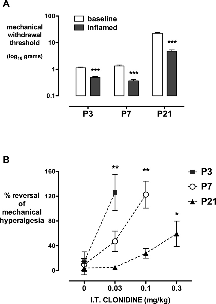
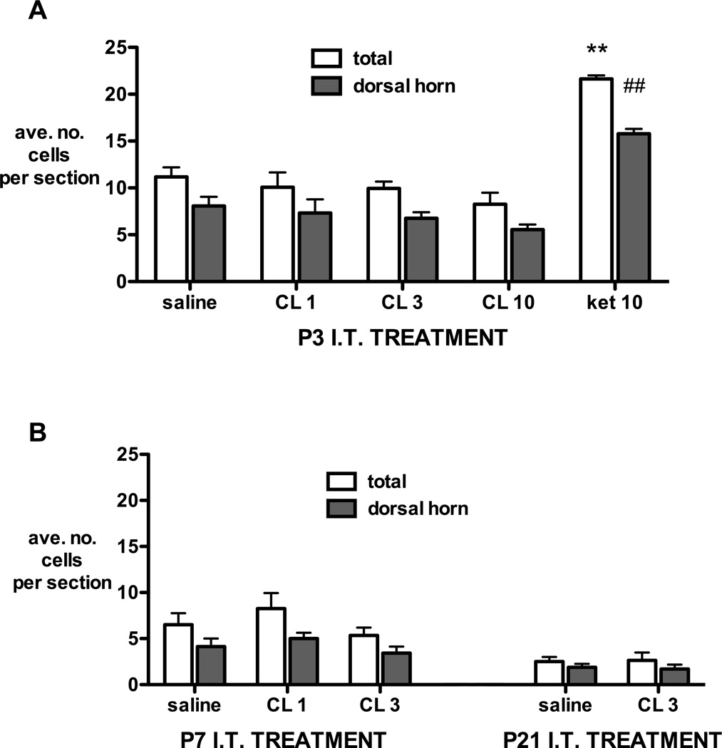
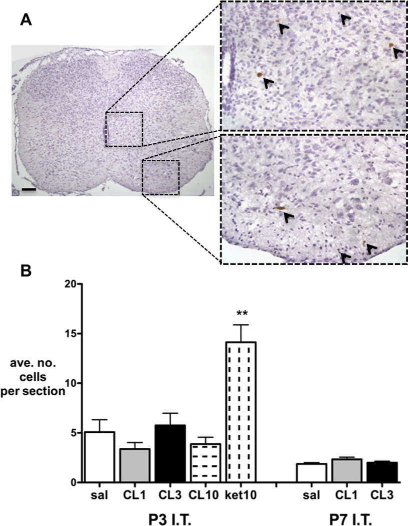
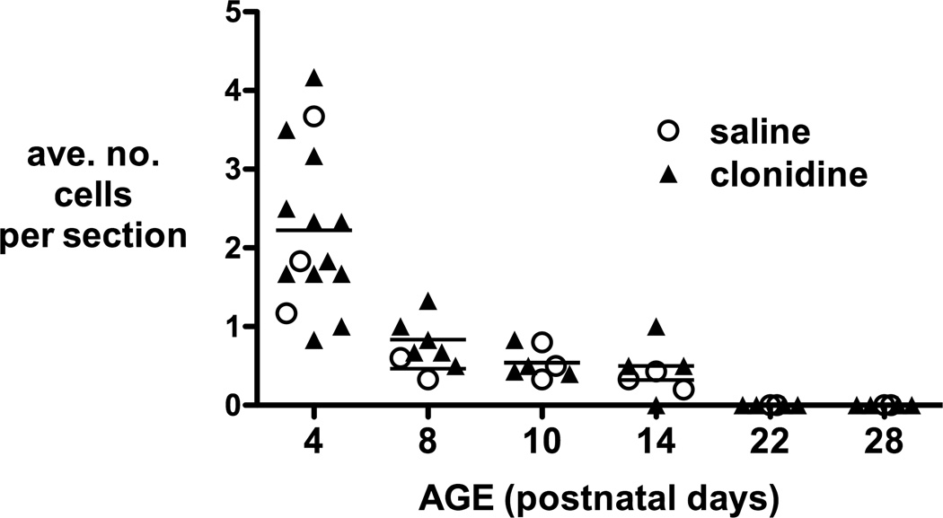
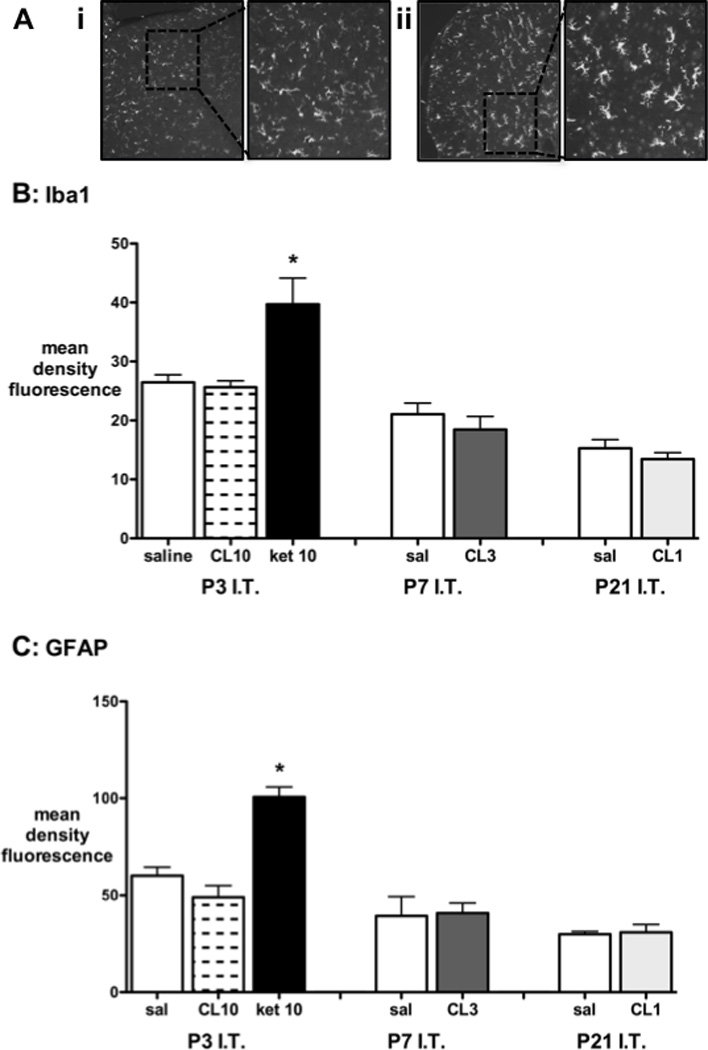
Methods: Rat pups aged 3, 7, or 21 postnatal (P) days were briefly anesthetized for intrathecal injections of saline or clonidine. At each age, the maximum tolerated, antinociceptive (increased hindlimb mechanical withdrawal threshold) and antihyperalgesic (hindpaw carrageenan inflammation) doses were determined. Lumbar spinal cord sections were assessed for apoptosis and cell death (histology, activated caspase-3 immunohistochemistry, Fluoro-Jade C staining), histopathology (hematoxylin and eosin staining), and increased glial reactivity (microglial and astrocytic markers). P3 intrathecal ketamine sections served as positive controls. In additional groups, thermal latency and mechanical withdrawal threshold were measured at P35.

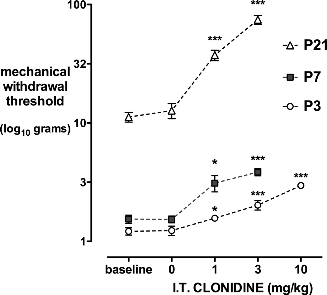
Results: Intrathecal clonidine produces age- and dose-dependent analgesia in rat pups. Maximal doses of clonidine did not alter the degree or distribution of apoptosis or increase glial reactivity in the neonatal spinal cord. No spinal histopathology was seen 1 or 7 days after injection at any age. Intrathecal clonidine did not produce persistent changes in reflex sensitivity to mechanical or thermal stimuli at P35.

Conclusions: Intrathecal clonidine in the postnatal rat did not produce signs of spinal cord toxicity, even at doses much larger than required for analgesia. The therapeutic ratio (maximum tolerated dose/antihyperalgesic dose) was >300 at P3, >30 at P7, and >10 at P21. These data provide additional information to inform the clinical choice of spinal analgesic drug in early life.


Conflict of interest statement

Conflicts: Suellen M. Walker reported no conflicts of interest

Conflicts: Marjorie Grafe reported no conflicts of interest

Conflicts: Tony L Yaksh reported no conflicts of interest

Figures

Figure 1
Figure 1
Dose-dependent anti-nociceptive effects of intrathecal clonidine. Mechanical withdrawal thresholds at baseline and 30 minutes following intrathecal (I.T.) injection of saline or clonidine (CL) 1, 3 or 10mg/kg in postnatal day (P) 3, 7 or 21 rat pups are shown. Data points = mean±SEM, n=6–8 per treatment group, *P<0.05 ***P<0.001 one way ANOVA with Dunnett’s comparison to baseline.
Figure 2
Figure 2
Anti-hyperalgesic effects of clonidine. A, Mechanical withdrawal thresholds are significantly reduced from baseline 3 hours following hindpaw carrageenan injection at postnatal day (P) 3, 7, and 21. Bars = mean±SEM, ***P<0.001 Student’s two-tailed paired t-test. B, The degree of reversal of carrageenan-induced hindpaw inflammatory hyperalgesia [30mins post dose – inflamed/baseline – inflamed) ×100] is shown following intrathecal (I.T.) saline (0) or increasing doses of intrathecal clonidine at postnatal (P) day 3, 7 and 21. Data=mean±SEM, n=5–7 per group, *P<0.05 **P<0.01 vs saline, one way ANOVA with Dunnett’s comparison to saline.
Figure 3
Figure 3
Fluoro-Jade C positive cell counts 24 hours following intrathecal injection at postnatal day 3 (A), 7 or 21 (B). The number of cells in the whole section (total) and the proportion distributed within the dorsal horn (dorsal horn) are averaged from at least 4 non-consecutive sections per animal. I.T.=intrathecal; P=postnatal day; CL=clonidine 1, 3 or 10mg/kg; ket 10 = ketamine 10mg/kg. Bars = mean±SEM, n= 4 animals per group. **P<0.01 ket 10 vs all groups in total section; ##P<0.01 ket 10 vs all groups in dorsal horn; one way ANOVA with Bonferroni post-hoc comparisons.
Figure 4
Figure 4
A, Example of lumbar spinal cord section following intrathecal ketamine at P3 with activated-caspase 3 immunopositive cells stained brown and highlighted with arrows. B, Activated-caspase 3 immunopositive cell counts 24 hours following intrathecal injection at postnatal day 3 or 7. Numbers are averaged from at least 4 non-consecutive sections per animal. I.T=intrathecal; P=postnatal day; CL=clonidine 1, 3 or 10mg/kg; ket=ketamine 10mg/kg. Bars = mean±SEM, n= 4 animals per group. **P<0.01 ket 10 vs all other P3 groups; one way ANOVA with Bonferroni post-hoc comparisons.
Figure 5
Figure 5
Apoptotic cell counts from haematoxylin and eosin stained sections. Numbers are averaged from at least 5 sections per animal 24 hours or 7 days following intrathecal injection on postnatal day 3, 7 and 21. Counts following intrathecal saline or any dose of intrathecal clonidine did not differ at any time point (n.s. one way ANOVA).
Figure 6
Figure 6
A, Representative spinal cord sections demonstrating Iba1 immunofluorescent staining 7 days following injection of i) intrathecal clonidine 10mg/kg or ii) intrathecal ketamine 10mg/kg. B, Quantification of Iba1 immunofluoresence in the dorsal horn 7 days following intrathecal (I.T.) saline, clondine 1, 3 or 10mg/kg (CL 1, 3, 10) or ketamine 10mg/kg (ket 10) at postnatal day (P)3, 7 or 21. C, Quantification of GFAP immunofluoresence in the dorsal horn in the same experimental groups as above. Bars=mean±SEM, n=3–4 animals per group. *P<0.05 ketamine 10mg/kg vs saline or clonidine 10mg/kg at P3; one way ANOVA with Bonferroni post-hoc comparisons.

References

    1. Reddy SV, Maderdrut JL, Yaksh TL. Spinal cord pharmacology of adrenergic agonist-mediated antinociception. J Pharmacol Exp Ther. 1980;213:525–533. - PubMed
    1. Yaksh TL, Reddy SV. Studies in the primate on the analgetic effects associated with intrathecal actions of opiates, alpha-adrenergic agonists and baclofen. Anesthesiology. 1981;54:451–467. - PubMed
    1. Yaksh TL. Pharmacology of spinal adrenergic systems which modulate spinal nociceptive processing. Pharmacol Biochem Behav. 1985;22:845–858. - PubMed
    1. Eisenach JC, De Kock M, Klimscha W. alpha(2)-adrenergic agonists for regional anesthesia. A clinical review of clonidine (1984–1995) Anesthesiology. 1996;85:655–674. - PubMed
    1. Eisenach JC, Lysak SZ, Viscomi CM. Epidural clonidine analgesia following surgery: phase I. Anesthesiology. 1989;71:640–646. - PubMed

Publication types

MeSH terms