

Thoracoscopic correction of a congenital persistent right aortic arch in a young cat
- PMID: 22467970
- PMCID: PMC3174512
Thoracoscopic correction of a congenital persistent right aortic arch in a young cat
Abstract
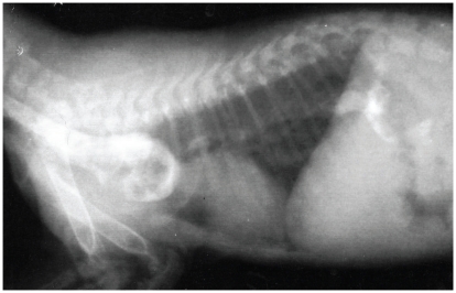
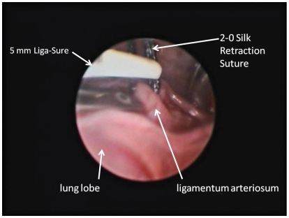
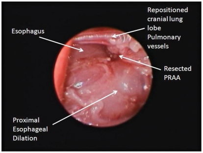
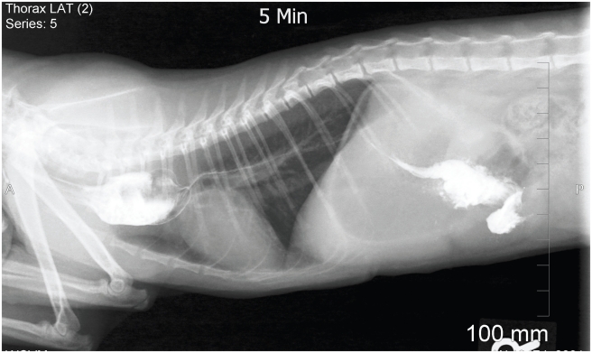
A 9-week-old kitten was diagnosed with a congenital vascular ring anomaly by means of an esophageal contrast study. At 6 mo of age, a non-selective vascular study was used to diagnose a persistent right aortic arch (PRAA). Left-sided thoracoscopic surgery was performed, using a Liga-Sure vessel sealant device to seal and transect the ligamentum arteriosum.
Correction par thoracoscopie d’une crosse aortique droite congénitale persistante chez un jeune chat. Un chaton âgé de 9 semaines a été diagnostiqué avec une anomalie congénitale d’anneau vasculaire à l’aide d’un examen œsophagien en contraste. À l’âge de 6 mois, une étude vasculaire non sélective a été utilisée pour diagnostiquer une crosse aortique droite persistante (PRAA). Une chirurgie par thorascopie du côté gauche a été réalisée à l’aide d’un dispositif de scellage des vaisseaux Liga-Sure pour sceller et couper transversalement le ligamentum arteriosum.
(Traduit par Isabelle Vallières)
Figures
References
-
- Kyles AE. Vascular ring anomalies. In: Slatter DG, editor. Textbook of Small Animal Surgery. 3rd ed. Philadelphia, Pennsylvania: Saunders; 2003. pp. 577–582.
-
- Berry AP, Brouwer GJ, Tennant BJ. Persistent right aortic arch in a kitten. Vet Rec. 1984;114:336–337. - PubMed
-
- Buchanan JW. Symposium. Thoracic surgery in the dog and cat — III: Patent ductus arteriosus and persistent right aortic arch surgery in dogs. J Small Anim Pract. 1968;9:409–428. - PubMed
-
- Jeffrey SC, Bataller N, Martin RA, Moon ML. Postsurgical nutritional management of megaesophagus secondary to persistent right aortic arch in a kitten. Feline Practice. 1995;23:17–23.
Publication types
MeSH terms
LinkOut - more resources
Full Text Sources
Miscellaneous