

Assessment of Bdellovibrio bacteriovorus 109J killing of Moraxella bovis in an in vitro model of infectious bovine keratoconjunctivitis
- PMID: 22468026
- PMCID: PMC3187635
Assessment of Bdellovibrio bacteriovorus 109J killing of Moraxella bovis in an in vitro model of infectious bovine keratoconjunctivitis
Abstract
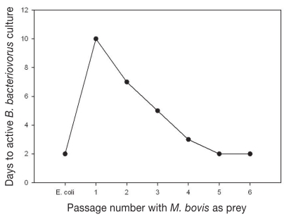
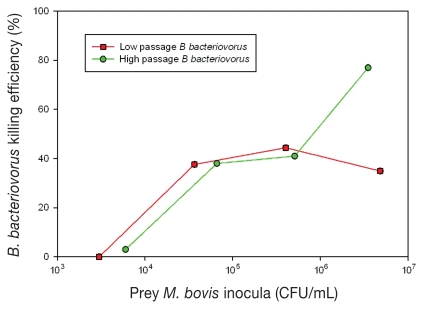
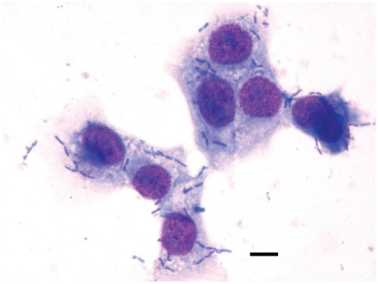
The objective of this study was to determine the potential of Bdellovibrio bacteriovorus 109J as an alternative non-chemotherapeutic treatment of infectious bovine keratoconjunctivitis (IBK). To accomplish this, various parameters of B. bacteriovorus predation of Moraxella bovis were determined in vitro. Initial passage of B. bacteriovorus using M. bovis as prey required 10 d for active cultures to develop compared with 2 d for culture on normal Escherichia coli prey; however by the 5th passage, time to active predatory morphology was reduced to 2 d. This high passage B. bacteriovorus culture [1 × 10(10) plaque forming units (PFU)/mL] killed 76% of M. bovis [1 × 10(7) colony forming units (CFU)/mL] present in suspension broth in a 4 h assay. The minimal level of M. bovis supporting B. bacteriovorus predation was 1 × 10(4) CFU/mL. To assess the ability of B. bacteriovorus to kill M. bovis on an epithelial surface mimicking IBK, an in vitro assay with Madin-Darby bovine kidney (MDBK) cells inoculated with 4 × 10(7) CFU/mL M. bovis was used. Treatment with a B. bacteriovorus suspension (1.6 × 10(11) PFU/mL) decreased adherence of M. bovis to MDBK cells by 6-fold at 12 h of treatment, as well as decreased the number of unattached M. bovis cells by 1.4-fold. This study demonstrates that B. bacteriovorus has potential as an effective biological control of M. bovis at levels likely present in IBK-infected corneal epithelia and ocular secretions.
Cette étude visait à déterminer le potentiel de Bdellovibrio bacteriovorus 109J comme traitement alternatif non-thérapeutique de la kérato-conjonctivite infectieuse bovine (IBK). À cet effet, divers paramètres de prédation de B. bacteriovorus envers Moraxella bovis ont été déterminés in vitro. Le premier passage de B. bacteriovorus utilisant M. bovis comme proie nécessitait 10 j pour qu’une culture active se développe comparativement à 2 j pour une culture utilisant Escherichia coli comme proie; toutefois, rendu au 5e passage, le temps requis pour obtenir la morphologie de prédateur actif était réduit à 2 j. Cette culture de passage élevé de B. bacteriovorus (1 × 10¹⁰ unités formatrices de plaques (PFU)/mL) a tué 76 % des M. bovis (1 × 10⁷ unités formatrices de colonies (CFU)/mL) présents dans un bouillon lors d’un essai d’une durée de 4 h. Le nombre minimal de M. bovis permettant la prédation par B. bacteriovorus était de 1 × 10⁴ CFU/mL. Afin d’évaluer la capacité de B. bacteriovorus à tuer M. bovis sur une surface épithéliale imitant IBK, une épreuve in vitro avec des cellules rénales bovines Madin-Darby (MDBK) inoculées avec 4 × 10⁷ CFU/mL M. bovis fut utilisée. Le traitement avec une suspension de B. bacteriovorus (1,6 × 10¹¹ PFU/mL) a réduit l’adhérence de M. bovis aux cellules MDBK par un facteur de 6 après 12 h de traitement, et a également diminué le nombre de cellules de M. bovis non-attachées par un facteur de 1,4. Cette étude démontre que B. bacteriovorus a le potentiel d’être un moyen de réduction biologique efficace de M. bovis à des niveaux susceptibles d’être présents sur l’épithélium cornéen et dans les sécrétions oculaires d’animaux infectés par l’IBK.
(Traduit par Docteur Serge Messier)
Figures
References
-
- Baptista PJHP. Infectious bovine keratoconjunctivitis: A review. Br Vet J. 1979;135:225–242. - PubMed
-
- George LW. Antibiotic treatment of infectious bovine keratoconjunctivitis. Cornell Vet. 1990;80:229–235. - PubMed
-
- Thrift FA, Overfield JR. Impact of pinkeye (infectious bovine keratoconjunctivitis) on weaning and postweaning performance of Hereford calves. J Ani Sci. 1974;38:1179–1184. - PubMed
-
- Brown MH, Brightman AH, Fenwick BW, et al. Infectious bovine keratoconjunctivitis: A review. J Vet Intern Med. 1998;12:259–266. - PubMed
-
- Smith JA, George LW. Treatment of acute ocular Moraxella bovis infections in calves with a parenterally administered long-acting oxytetracycline formulation. Am J Vet Res. 1985;46:804–807. - PubMed
Publication types
MeSH terms
Grants and funding
LinkOut - more resources
Full Text Sources
Other Literature Sources