Giant cell tumor of the mandible
- PMID: 22468203
- PMCID: PMC3314806
- DOI: 10.3342/ceo.2012.5.1.49
Giant cell tumor of the mandible
Abstract
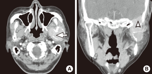
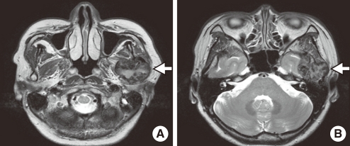
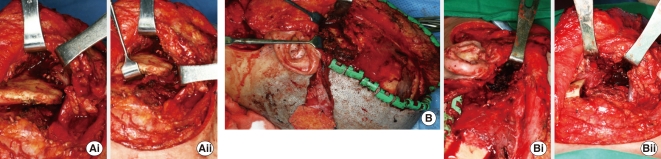
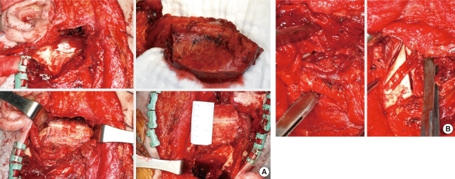
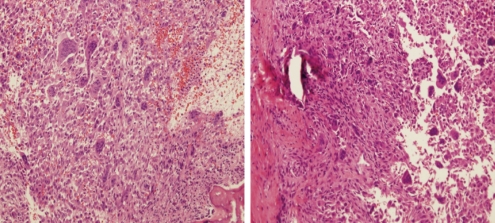
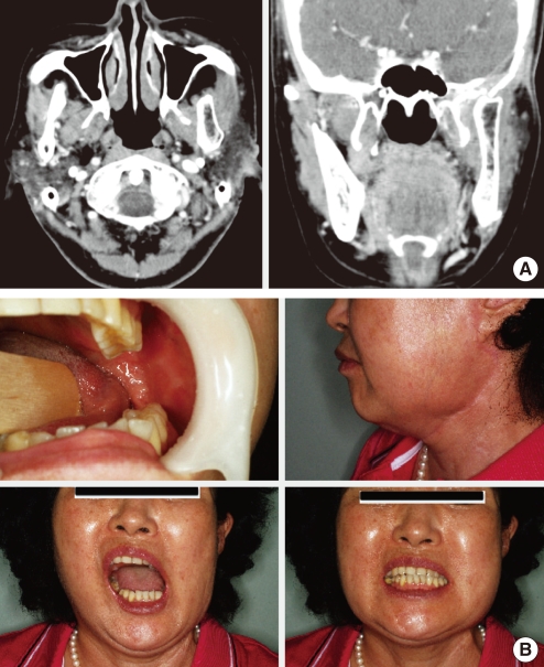
A 53-year-old woman presented with left mandibular area pain, trismus, and facial numbness that had persisted for 4 years. Physical examination revealed a 3×5 cm, hard, non-tender, and round mass on the left mandibular area. Computed tomography and magnetic resonance imaging revealed an expansile tumor involving the left mandibular ramus and temporomandibular joint area with bone destruction, extending to the base of middle cranial fossa and left zygomatic bone. The mass at the segment of left mandible and zygomatic bone, and base of middle cranial fossa was removed. Pathological examination of the mass revealed a giant cell tumor. The defect was reconstructed with iliac bone for the mandible and temporal bone and fascia for the cranial bone and dura. The case is described along with a review of the literature.
Keywords: Giant cell tumor; Mandible; Skull.
Conflict of interest statement
No potential conflict of interest relevant to this article was reported.
Figures
References
-
- Dahlin DC, Cupps RE, Johnson EW., Jr Giant-cell tumor: a study of 195 cases. Cancer. 1970 May;25(5):1061–1070. - PubMed
-
- Marcove RC, Lyden JP, Huvos AG, Bullough PB. Proceedings: Giant cell tumors treated by cryosurgery: an analysis of 25 cases. Proc Natl Cancer Conf. 1972;7:951–957. - PubMed
-
- Marcove RC, Lyden JP, Huvos AG, Bullough PB. Giant-cell tumors treated by cryosurgery: a report of twenty-five cases. J Bone Joint Surg Am. 1973 Dec;55(8):1633–1644. - PubMed
-
- Saleh EA, Taibah AK, Naguib M, Aristegui M, Vassallo G, Landolfi M, et al. Giant cell tumor of the lateral skull base: a case report. Otolaryngol Head Neck Surg. 1994 Sep;111(3 Pt 1):314–318. - PubMed
-
- Rosenbloom JS, Storper IS, Aviv JE, Hacein-Bey L, Bruce JN. Giant cell tumors of the jugular foramen. Am J Otolaryngol. 1999 May-Jun;20(3):176–179. - PubMed