. 2012 Mar;83(5):953-67.
doi: 10.1111/j.1365-2958.2012.07980.x.

Characterization of the Group A Streptococcus Mga virulence regulator reveals a role for the C-terminal region in oligomerization and transcriptional activation

Affiliations

Characterization of the Group A Streptococcus Mga virulence regulator reveals a role for the C-terminal region in oligomerization and transcriptional activation

Elise R Hondorp et al. Mol Microbiol. 2012 Mar.

Abstract

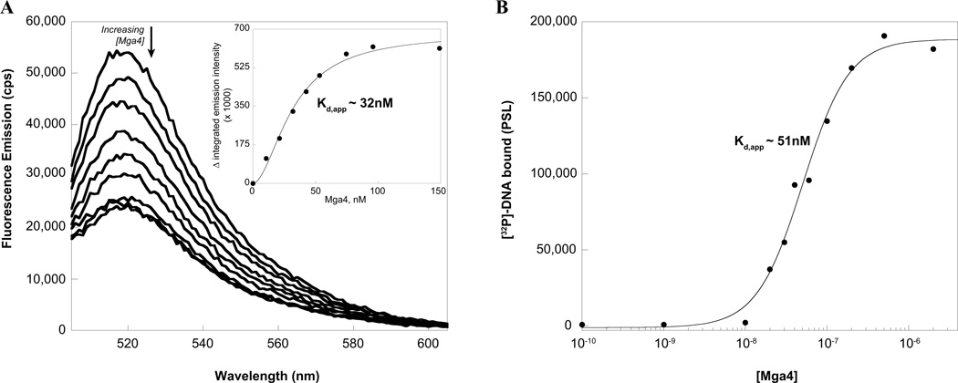
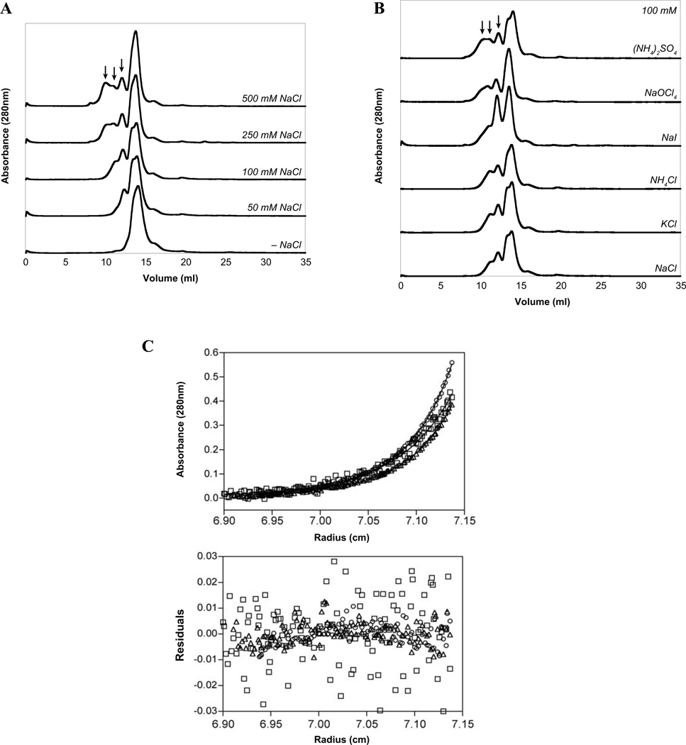
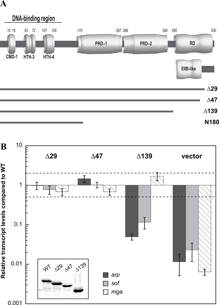
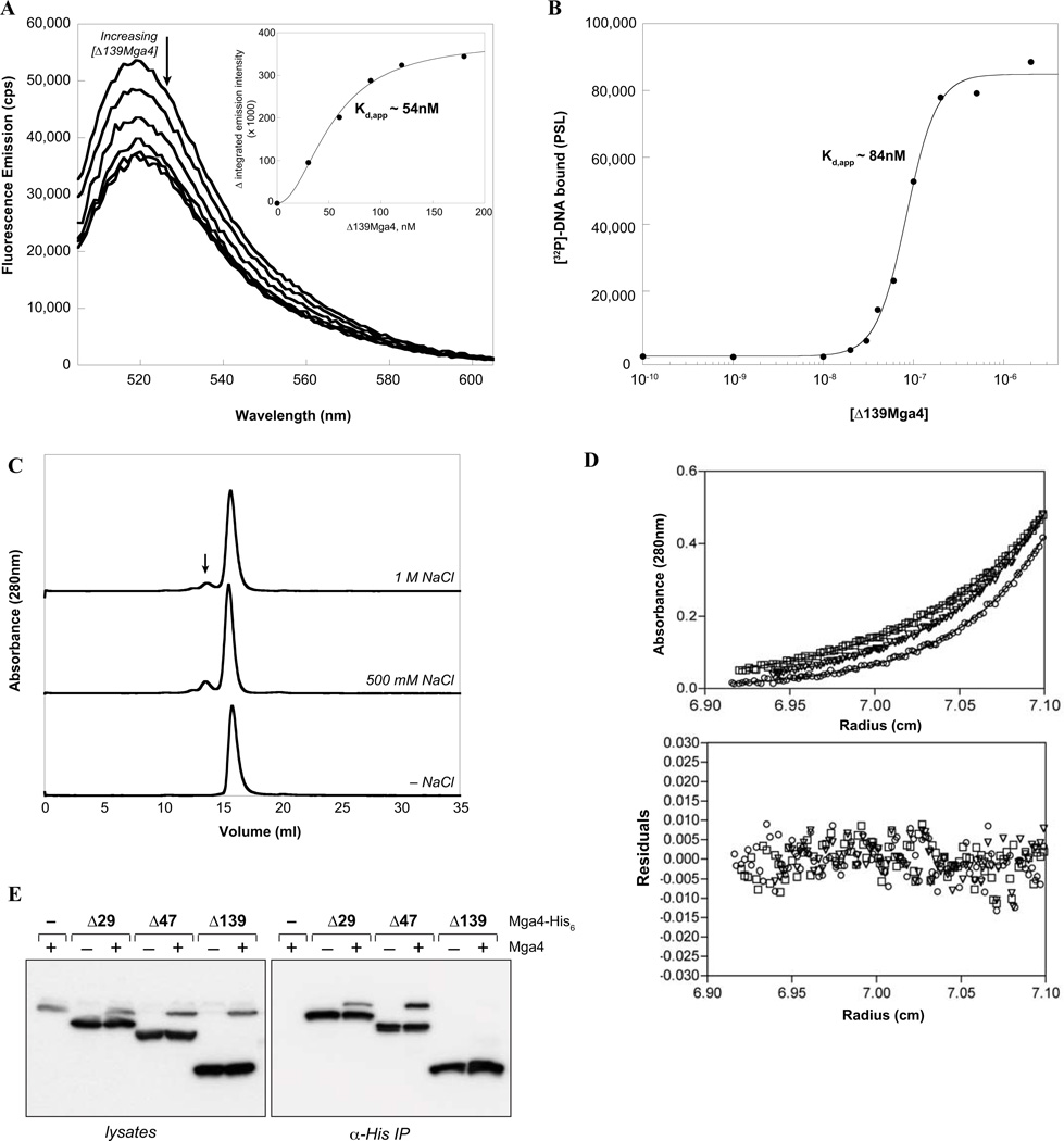
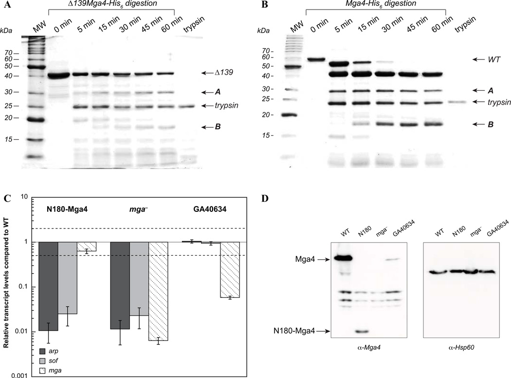
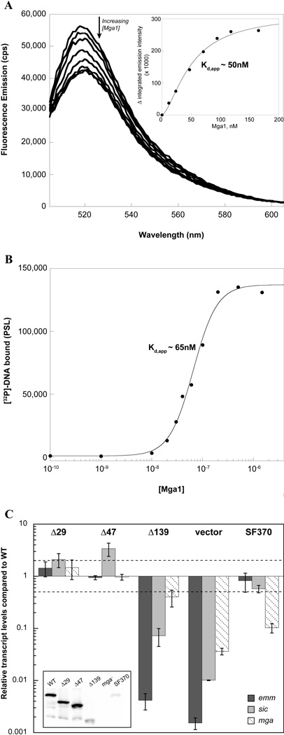
The Group A Streptococcus (GAS) is a strict human pathogen that causes a broad spectrum of illnesses. One of the key regulators of virulence in GAS is the transcriptional activator Mga, which co-ordinates the early stages of infection. Although the targets of Mga have been well characterized, basic biochemical analyses have been limited due to difficulties in obtaining purified protein. In this study, high-level purification of soluble Mga was achieved, enabling the first detailed characterization of the protein. Fluorescence titrations coupled with filter-binding assays indicate that Mga binds cognate DNA with nanomolar affinity. Gel filtration analyses, analytical ultracentrifugation and co-immunoprecipitation experiments demonstrate that Mga forms oligomers in solution.Moreover, the ability of the protein to oligomerize in solution was found to correlate with transcriptional activation; DNA binding appears to be necessary but insufficient for full activity. Truncation analyses reveal that the uncharacterized C-terminal region of Mga, possessing similarity to phosphotransferase system EIIB proteins, plays a critical role in oligomerization and in vivo activity. Mga from a divergent serotype was found to behave similarly, suggesting that this study describes a general mechanism for Mga regulation of target virulence genes within GAS and provides insight into related regulators in other Gram-positive pathogens.


Figures

FIG. 1
FIG. 1. Mga4-His6 binds to Parp4 in vitro
A, Fluorescence titration. The fluorescence spectrum of a FAM-labeled 49mer containing the Mga-binding site of the Parp4 promoter was monitored upon the addition of purified Mga4-His6 to assess binding. The decrease in integrated emission intensity from 515 to 540 nm was plotted versus the concentration of Mga4-His6 to determine the apparent Kd for half maximal binding (inset). B, Filter binding assays. Varying concentrations of Mga4-His6 were incubated with the [32P]-labeled Parp4 49mer and then filtered through nitrocellulose, which retains the protein-bound radiolabel. Phosphorimager analysis followed by densitometry was used to plot the amount of DNA bound as a function of Mga4-His6 concentration and determine Kd,app
FIG. 2
FIG. 2. Mga forms oligomers in solution
Mga4-His6 was incubated in a HEPES/Citrate buffer containing varying salts and then subjected to gel filtration analysis in the same buffer. A, Increasing the concentration of NaCl results in the appearance of higher-order multimers (indicated by arrows). B, Maintaining the salt concentration at 100 mM while varying the identity of the anion (but not the cation) also impacts the oligomerization pattern of Mga4-His6. C, Mga4-His6 in the presence of 100 mM NaCl was analyzed by sedimentation equilibrium analytical ultracentrifugation for various protein concentrations (7.5 µM, circles; 20 µM, squares; and 30 µM, triangles) centrifuged at 16,000 rpm. Fifteen datasets were fit to a monomer-dimer model (solid lines) resulting in the residuals depicted.
FIG. 3
FIG. 3. The C-terminal region of Mga4-His6 is required for in vivo activity
A, Schematic showing N-terminal regions implicated in DNA binding (CMD-1, HTH-3, and HTH-4) and central domains with strong structural homology to PTS regulatory domains (PRD-1 and PRD-2). The C-terminal region at one time predicted to be a CheY-like receiver domain (RD) is indicated. Newly recognized structural homology to an EIIB domain is depicted below the RD. Lines representing the different C-terminal truncation mutants are shown. B, Full-length and truncated Mga4-His6 proteins (Δ29Mga4-His6, Δ47Mga4-His6, and Δ139Mga4-His6) were each expressed on a plasmid under the native Pmga4 promoter in a mga4 strain, KSM547. Mga activity was assayed in vivo by real-time RT-PCR analysis of the Mga-regulated genes, arp (dark gray), sof (light gray), and mga itself (diagonal hashes). Relative transcript levels for the mutant strains compared to the wild type are presented. Error bars represent the standard error from six biological replicates. Differences greater than 2-fold in expression for the mutant strains compared to wild type (denoted by a dashed line) were considered significant. Empty vector mga results are shown alongside for comparison. The western blot of cell lysates probed with the α-Mga4 antibody (inset) is shown.
FIG. 4
FIG. 4. Δ139Mga4-His6 binds DNA with an affinity similar to full-length Mga, but is defective for oligomerization
A, Fluorescence titration. The fluorescence spectrum of a FAM-labeled 49mer containing the Mga-binding site of the Parp4 promoter was monitored upon the addition of purified Δ139Mga4-His6 to assess binding. The decrease in integrated emission intensity from 515 to 540 nm was plotted versus the concentration of Δ139Mga4-His6 to determine the apparent Kd (inset). B, Filter binding assays. Varying concentrations of Δ139Mga4-His6 were incubated with the [32P]-labeled Parp4 49mer. The complex was filtered through nitrocellulose to assess the amount of DNA bound as a function of Δ139Mga4-His6 concentration and determine Kd,app. C, Gel filtration analysis. Δ139Mga4-His6 was incubated in a HEPES/Citrate buffer containing varying concentrations of NaCl and then subjected to size exclusion chromatography. An arrow indicates the position of a higher-order multimer upon increasing salt concentrations. D, Sedimentation equilibrium by analytical ultracentrifugation. Δ139Mga4-His6 (7.5 µM) in the presence of 100 mM NaCl was centrifuged at 18,000 (squares), 20,000 (inverted triangles) and 22,000 (circles) rpm. Six datasets were fit to a model for a single homogenous species (solid lines) resulting in the residuals depicted. E. Co-immunoprecipitation of Mga4 with truncated proteins. Δ29-, Δ47-, and Δ139Mga4-His6 proteins were expressed in wild type (GA40634) and mga4 (KSM547) GAS strains. His-tagged proteins were immunoprecipitated using α-His, and coimmunoprecipitation of endogenous Mga4 was probed by western blot analysis with α-Mga4 following separation of proteins by SDS-PAGE (right). Similar western blots of whole lysates prior to immunoprecipitation are shown to verify protein expression (left).
FIG. 5
FIG. 5. The N-terminal DNA-binding region of Mga is not active in vivo
A & B, Limited tryptic digestion of native Δ139Mga4-His6 (A) and full-length Mga4-His6 (B) with trypsin (0.12% or 0.08%, respectively) followed over time by SDS-PAGE. The predominant fragments observed over extended digestions (denoted as A and B) are indicated. C, In vivo activity of N180-Mga4. His-tagged wild-type and N180-Mga4 proteins were expressed from the native Pmga4 promoter in a mga4 strain, KSM547. Mga activity was assayed by real-time RT-PCR of the Mga-regulated genes, arp (dark gray), sof (light gray), and mga (diagonal hashes). Relative transcript levels for the mutant strains compared to the wild type are presented. Error bars represent the standard error from three biological replicates. Differences greater than 2-fold (denoted by a dashed line) in expression for the mutant strains compared to wild type were considered significant. The empty vector results in the mga and wild-type strain are shown for comparison. D, Western blot analyses. Immunoblots of cell lysates probed with α-Mga4 (left) reveal the levels of full-length and N180-Mga4 proteins (indicated by arrows) in a mga4 strain, KSM547 expressing either Mga4-His6 (WT), N180-His6 (N180) or an empty vector (mga ), and an isogenic parental M4 (GA40634) strain. An α-Hsp60 blot (right) was performed to demonstrate equal loading.
FIG. 6
FIG. 6. A mga-1 Mga behaves similarly to a mga-2 Mga
A, Fluorescence titration. The fluorescence spectrum of a FAM-labeled 49mer containing the Mga-binding site of Parp4 was monitored upon the addition of purified Mga1-His6 to assess binding. The decrease in integrated emission intensity from 515 to 540 nm was plotted versus the concentration of Mga1-His6 to determine the apparent Kd (inset). B, Filter binding assays. Varying concentrations of Mga1-His6 were assayed with the [32P]-labeled Parp4 49mer. The complex was filtered through nitrocellulose to assess the amount of DNA bound as a function of Mga1-His6 concentration and determine Kd,app. C, In vivo activity of M1 truncated proteins. Full-length and truncated Mga1-His6 proteins (Δ29Mga1-His6, Δ47Mga1-His6, and Δ139Mga1-His6) were each expressed from the native Pmga1 promoter in a mga1 strain, KSM1656L. Mga activity was assayed by real-time RT-PCR of the Mga1-regulated genes, emm (dark gray), sic (light gray), and mga (diagonal hashes). Relative transcript levels for the mutant strains compared to the wild type are presented. Error bars represent the standard error from three biological replicates. Differences greater than 2-fold (denoted by a dashed line) in expression for the mutant strains compared to wild type were considered significant. Results for the empty vector in the mga strain (vector) as well as endogenous Mga1 in the isogenic wild type strain (SF370) are shown alongside for comparison. A western blot of cell lysates probed with α-Mga1 (inset) is shown.

References

    1. Almengor AC, McIver KS. Transcriptional activation of sclA by Mga requires a distal binding site in Streptococcus pyogenes. J Bacteriol. 2004;186:7847–7857. - PMC - PubMed
    1. Almengor AC, Walters MS, McIver KS. Mga is sufficient to activate transcription in vitro of sof/sfbX and other Mga-regulated virulence genes in the group A streptococcus. J Bacteriol. 2006;188:2038–2047. - PMC - PubMed
    1. Andersson G, McIver K, Heden LO, Scott JR. Complementation of divergent mga genes in group A streptococcus. Gene. 1996;175:77–81. - PubMed
    1. Aravind L, Anantharaman V, Balaji S, Babu MM, Iyer LM. The many faces of the helix-turn-helix domain: transcription regulation and beyond. FEMS Microbiol Rev. 2005;29:231–262. - PubMed
    1. Bessen DE, Manoharan A, Luo F, Wertz JE, Robinson DA. Evolution of transcription regulatory genes is linked to niche specialization in the bacterial pathogen Streptococcus pyogenes. J Bacteriol. 2005;187:4163–4172. - PMC - PubMed

MeSH terms