. 2012 Apr 24;51(16):3383-93.
doi: 10.1021/bi300007r. Epub 2012 Apr 10.

Solution structural ensembles of substrate-free cytochrome P450(cam)

Affiliations

Solution structural ensembles of substrate-free cytochrome P450(cam)

Eliana K Asciutto et al. Biochemistry. .

Abstract

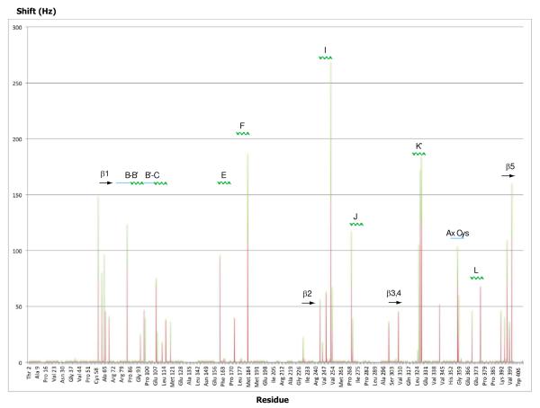
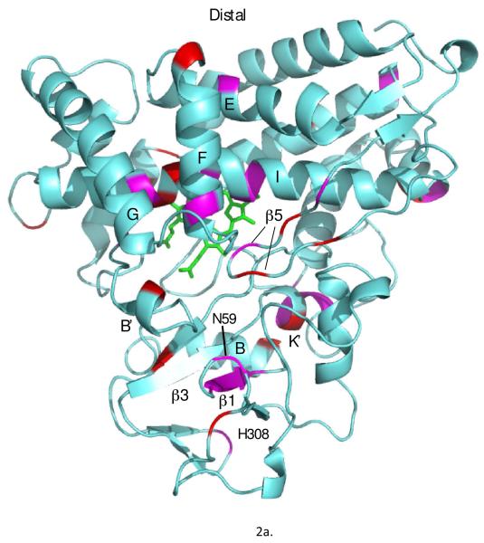
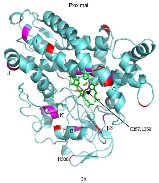
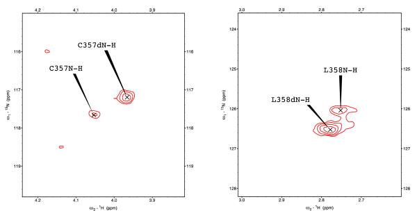
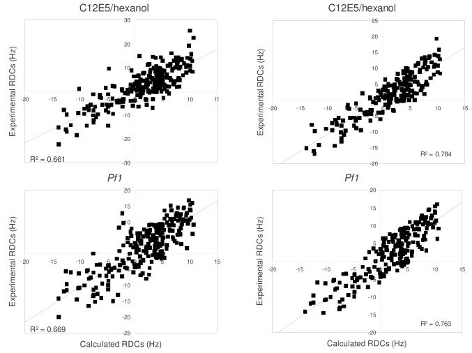
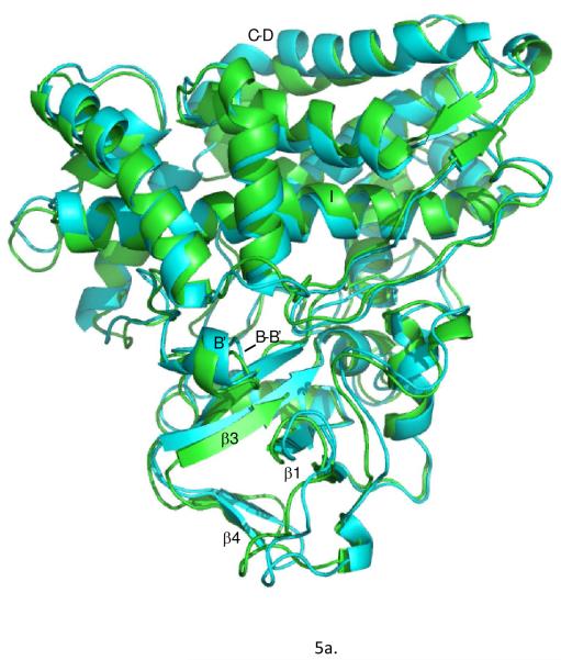
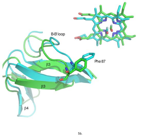
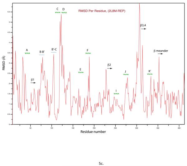
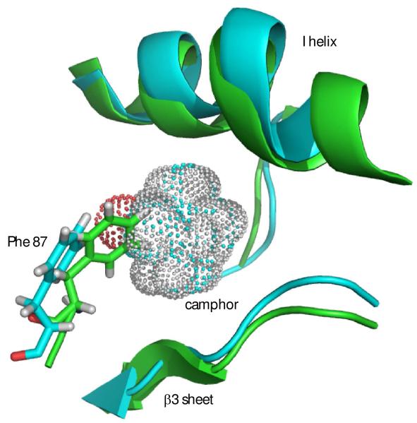
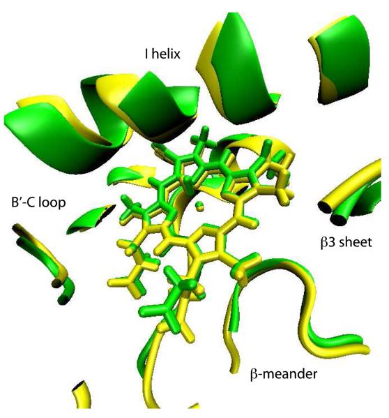
Removal of substrate (+)-camphor from the active site of cytochrome P450(cam) (CYP101A1) results in nuclear magnetic resonance-detected perturbations in multiple regions of the enzyme. The (1)H-(15)N correlation map of substrate-free diamagnetic Fe(II) CO-bound CYP101A permits these perturbations to be mapped onto the solution structure of the enzyme. Residual dipolar couplings (RDCs) were measured for (15)N-(1)H amide pairs in two independent alignment media for the substrate-free enzyme and used as restraints in solvated molecular dynamics (MD) simulations to generate an ensemble of best-fit structures of the substrate-free enzyme in solution. Nuclear magnetic resonance-detected chemical shift perturbations reflect changes in the electronic environment of the NH pairs, such as hydrogen bonding and ring current shifts, and are observed for residues in the active site as well as in hinge regions between secondary structural features. RDCs provide information about relative orientations of secondary structures, and RDC-restrained MD simulations indicate that portions of a β-rich region adjacent to the active site shift so as to partially occupy the vacancy left by removal of the substrate. The accessible volume of the active site is reduced in the substrate-free enzyme relative to the substrate-bound structure calculated using the same methods. Both symmetric and asymmetric broadening of multiple resonances observed upon substrate removal as well as localized increased errors in RDC fits suggest that an ensemble of enzyme conformations are present in the substrate-free form.


Figures

Figure 1
Figure 1
Chemical shift perturbations observed in 1H,15N TROSY-HSQC spectra upon removal of substrate from reduced and CO-bound CYP101A1 displayed as a function of sequence and secondary structure. Both 1H (orange bars) and 15N (green bars) shifts are shown, so the y-axis value is the sum of perturbations in sec-1 (Hz) observed in both dimensions for a given correlation. Secondary structural features with which perturbed residues are associated are indicated above, with solid arrow representing β structure, zig-zag green lines for helix, and straight blue lines for loop or irregular structure. Secondary structural features are named using the conventions of Poulos et al. (21). The Ax Cys label refers to Cys 357 and Leu 358.
Figure 2
Figure 2
Chemical shift perturbations observed in 1H,15N TROSY-HSQC spectra upon removal of substrate from reduced and CO-bound CYP101A1 superimposed on the solution structure 2L8M (ref. (14)). Only those residues for which N-H correlations distinct from those of the substrate-bound form are observed are highlighted. Residues highlighted in magenta exhibit shifts > 40 Hz in either the 1H or 15N dimensions, while smaller shifts are represented in red. Top: distal view (active site cavity towards the reader) with perturbed secondary structural features or residue IDs labeled using the conventions of Poulos et al. (21). Bottom: Proximal view, rotated slightly for clarity.
Figure 2
Figure 2
Chemical shift perturbations observed in 1H,15N TROSY-HSQC spectra upon removal of substrate from reduced and CO-bound CYP101A1 superimposed on the solution structure 2L8M (ref. (14)). Only those residues for which N-H correlations distinct from those of the substrate-bound form are observed are highlighted. Residues highlighted in magenta exhibit shifts > 40 Hz in either the 1H or 15N dimensions, while smaller shifts are represented in red. Top: distal view (active site cavity towards the reader) with perturbed secondary structural features or residue IDs labeled using the conventions of Poulos et al. (21). Bottom: Proximal view, rotated slightly for clarity.
Figure 3
Figure 3
Chemical shift perturbations observed in 1H,15N TROSY-HSQC spectra in camphor-depleted samples of reduced CO-bound CYP101A1 for heme axial cysteine ligand C357 (left) and adjacent L358 (right). Correlations marked C357N-H and L358N-H are at the positions observed for camphor-bound CYP101A1 and are assigned to residual camphor-bound enzyme. Those peaks marked as C357dN-H and L358dN-H are shifted relative to their positions in camphor-bound enzyme and are assigned to the camphor-free form.
Figure 4
Figure 4
Fitting of experimental RDCs versus RDC values calculated from MD simulations. Top left, fitting of all 228 RDCs measured in C12E5/hexanol medium used in MD simulations. Top right, fitting after removal of outliers with deviations > 6 Hz from calculated values (see text). All RDC values are in sec-1 (Hz). Bottom left, fitting of all 228 RDCs measured in Pf1 phage medium used in MD simulations. Bottom right, fitting after removal of outliers with deviations > 6 Hz from calculated values (see text).
Figure 5
Figure 5
a) Substrate-bound 2L8M structure (green) aligned by Cα carbon on REP1 (cyan), identified as the substrate-free structure with lowest RDC restraint violation energy. b) Close-up view of the same superposition highlighting the displacements of the heme, the B-B’ loop including Phe 87 and the β3-β4 domain. RMSD values between the two structures per residue are shown in the Figure 5c.
Figure 5
Figure 5
a) Substrate-bound 2L8M structure (green) aligned by Cα carbon on REP1 (cyan), identified as the substrate-free structure with lowest RDC restraint violation energy. b) Close-up view of the same superposition highlighting the displacements of the heme, the B-B’ loop including Phe 87 and the β3-β4 domain. RMSD values between the two structures per residue are shown in the Figure 5c.
Figure 6
Figure 6
Contraction of active site in REP1 (green) from best-fit backbone alignment to 2L8M (cyan). Camphor from 2L8M is shown as a van der Waals dot surface. Camphor-bound 2L8M structure is shown in cyan. Note that the side chain of Phe 87 partially occupies volume vacated by camphor. The side chains of Leu 244 and Val 247 also partially occlude the space occupied by camphor (see text). Unless otherwise noted, structural figures are generated using PyMOL (35).
Figure 7
Figure 7
2L8M (green) and REP1 (yellow) structures aligned using the heme macrocycle as the reference. Residues at distances less than 6 Å from the heme are shown using a cartoon representation.

References

    1. Fischer E. Einfluss der Configuration auf die Wirkung der Enzyme. Ber. Deutsche Chem. Gessel. 1894;27:2985–2993.
    1. Koshland DE. Application of a theory of enzyme specificity to protein synthesis. Proc. Natl. Acad. Sci. USA. 1958;44:98–104. - PMC - PubMed
    1. Niesen MJM, Bhattacharya S, Vaidehi N. The role of conformational ensembles in ligand recognition in G-protein coupled receptors. J. Am. Chem. Soc. 2011;133:13197–13204. - PubMed
    1. Fraser JS, van den Bedem H, Samelson AJ, Lang PT, Holton JM, Echols N, Alber T. Accessing protein conformational ensembles using room-temperature X-ray crystallography. Proc. Natl. Acad. Sci. USA. 2011;108:16247–16252. - PMC - PubMed
    1. Frauenfelder H, Petsko GA, Tsernoglou D. Temperature-dependent X-ray diffraction as a probe of protein structural dynamics. Nature. 1979;280:558–563. - PubMed

Publication types

Associated data