. 2012 Sep 20;21(14):2703-17.
doi: 10.1089/scd.2011.0604. Epub 2012 May 17.

Hypoxia-inducible factor 1-α-AA-modified bone marrow stem cells protect PC12 cells from hypoxia-induced apoptosis, partially through VEGF/PI3K/Akt/FoxO1 pathway

Affiliations

Hypoxia-inducible factor 1-α-AA-modified bone marrow stem cells protect PC12 cells from hypoxia-induced apoptosis, partially through VEGF/PI3K/Akt/FoxO1 pathway

Qian Zhong et al. Stem Cells Dev. .

Abstract

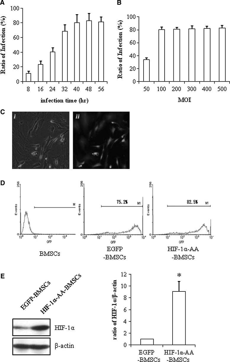
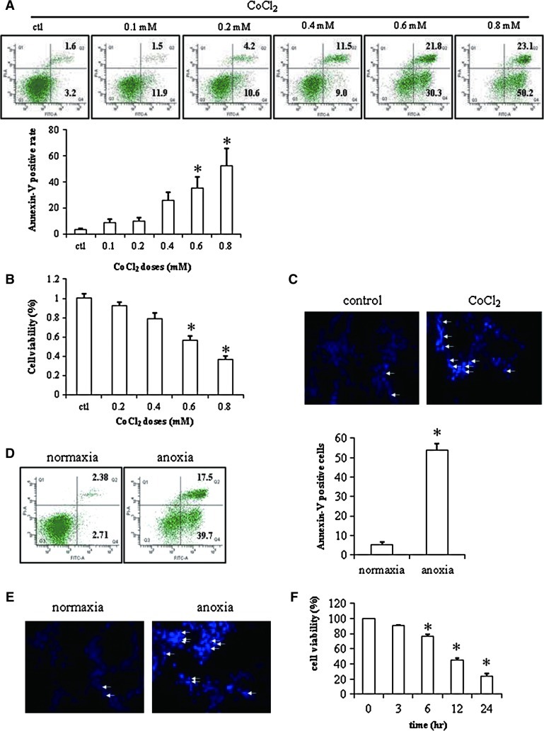
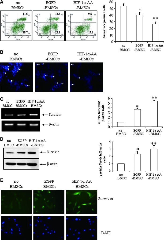
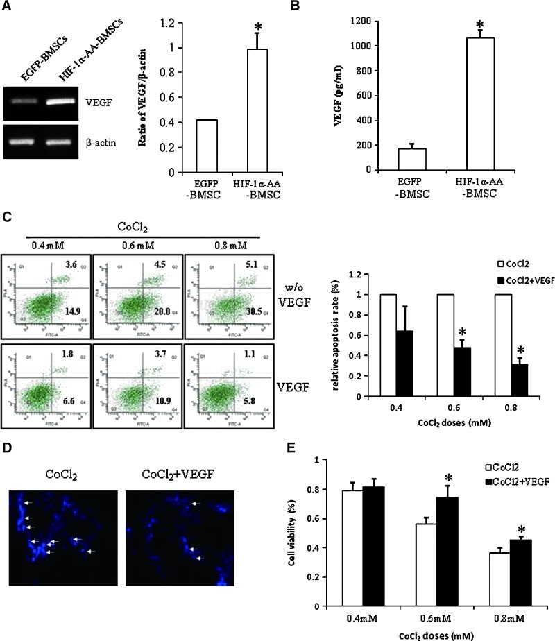
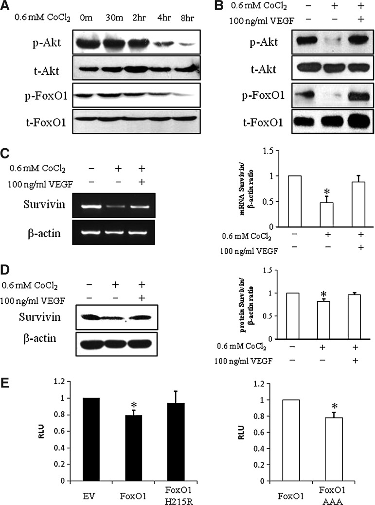
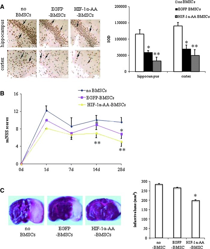
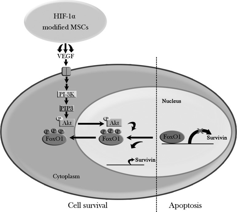
Bone marrow stem cells (BMSCs) have been shown to improve neurological function recovery in cerebral ischemia. Hypoxia-inducible factor-1 (HIF-1) α-AA is a more stable mutant form of HIF-1α, which is a crucial oxygen-sensitive regulator. To investigate the protective effects of HIF-1α-AA-modified BMSCs on neuron survival in cerebral ischemia models, we co-cultured HIF-1α-AA-modified BMSCs with neuron-like cells (PC12 cells) and observed a significant increase in the release of vascular endothelial growth factor (VEGF) from BMSCs, the decreased PC12 cell apoptosis, and the upregulation of Survivin expression reduced by hypoxia in PC12 cells compared to enhanced green fluorescent protein (EGFP) BMSCs. In addition, to explore whether VEGF secreted by HIF-1α-AA-modified BMSCs plays an important role in preventing hypoxia-induced apoptosis and the possible mechanism involved, exogenous VEGF were applied and the similar protective effects on PC12 cells were observed in vitro. Furthermore, hypoxia reduced the expression of phosphorylated Akt and phosphorylated FoxO1, whereas the administration of VEGF reversed these changes. Transfection of FoxO1 H215R, a DNA-binding mutant, abrogated the inhibitory ability on Survivin promoter activity, whereas FoxO1 AAA, the active form of FoxO1, presented further repression on Survivin promoter, indicating that FoxO1 directly binds on Survivin promoter as a transcriptional repressor and that phosphorylation status of FoxO1 affects its inhibition on the Survivin promoter. Transplantation of HIF-1α-AA-modified BMSCs after cerebral ischemia in vivo sufficiently reduced neurons apoptosis, decreased cerebral infarction volume, and induced a significant improvement on the modified neurological severity score compared to the EGFP BMSCs group. In conclusion, HIF-1α-AA-modified MSCs showed an obvious protective effect on neuron-like cells or neuron after ischemia in vitro and in vivo, at least in part, through the VEGF/PI3K/Akt/FoxO1 pathway.


Figures

FIG. 1.
FIG. 1.
Establishment of lentiviral-mediated hypoxia-inducible factor (HIF)-1α-AA-modified bone marrow stem cells (BMSCs). (A) Flow cytometry analysis of green fluorescent protein (GFP) expression in BMSCs infected with Lv-enhanced green fluorescent protein (EGFP) at different time points when Multiplicity of Infection (MOI)=100. (B) Flow cytometry analysis of GFP expression in BMSCs infected with Lv-EGFP at different MOI after 48 h. (C) Representative pictures of both phase image and fluorescein isothiocyanate image of HIF1α-AA BMSCs (100×). (D) Flow cytometry analysis of GFP expression in BMSCs, EGFP-BMSCs, and HIF1α-AA BMSCs at MOI=100. (E) Representative picture of western blotting of HIF1α expression in EGFP-BMSCs, and HIF1α-AA BMSCs. β-actin was used as a control for protein loading. P≤0.05; data are means±SEM (n=3); *significant difference from EGFP-BMSCs.
FIG. 2.
FIG. 2.
Apoptosis of PC12 cells. (A) Histograms and quantitative analysis of flow cytometry of Annexin-V/propidium iodide (PI) staining in PC12 cells treated with different doses of CoCl2 (n=3). (B) MTT assay of PC12 cells treated with different doses of CoCl2 (n=8). (C) Hoechst staining of PC12 cells treated with or without 0.6 mM CoCl2 (100×) (n=2). (D) Histograms and quantitative analysis of flow cytometry of Annexin-V/PI staining in PC12 cells under normoxic or anoxic condition for 12 h (n=3). (E) Hoechst staining of PC12 cells under normoxic or anoxic condition for 12 h (100×) (n=2). Hoechst-positive staining is denoted by white arrows. (F) MTT assay of PC12 cells under anoxic condition at different time points (n=3). Data are means±SEM; P≤0.05; *significant difference from control. Color images available online at www.liebertonline.com/scd
FIG. 3.
FIG. 3.
HIF1α-AA-modified BMSCs protect PC12 cells from apoptosis. (A) Histograms and quantitative analysis of flow cytometry of Annexin-V/PI staining in PC12 cells under anoxic condition for 12 h without co-culturing of BMSCs (no BMSCs), or with co-culturing of EGFP-BMSCs or HIF1α-AA-BMSCs (n=3). (B) Hoechst staining of PC12 cells under anoxic condition for 12 h without co-culturing of BMSCs (no BMSCs), or with co-culturing of EGFP-BMSCs or HIF1α-AA-BMSCs (100×) (n=2). Polymerase chain reaction (PCR) (n=3) (C), western blotting (n=3) (D), and immunostaining (n=2) of Survivin (green) (E) in PC12 cells under anoxic condition for 12 h without co-culturing of BMSCs (no BMSCs), or with co-culturing of EGFP-BMSCs or HIF1α-AA-BMSCs (400×). Nuclei were counterstained with 4′,6-diamidino-2-phenylindole. Data are means±SEM; P≤0.05; *significant difference from no BMSCs; **significant difference from other conditions. Color images available online at www.liebertonline.com/scd
FIG. 4.
FIG. 4.
Vascular endothelial growth factor (VEGF) secreted from HIF1α-AA-modified BMSCs prevents apoptosis. (A) Representative picture of PCR of VEGF expression in EGFP BMSCs or HIF1α-AA-BMSCs. β-actin was used as a control for protein loading. Data are means±SEM (n=3); P≤0.05; *significant difference from EGFP BMSCs. (B) The level of the released VEGF from EGFP BMSCs or HIF1α-AA-BMSCs by ELISA. Data are means±SEM (n=4); P≤0.05; *significant difference from EGFP BMSCs. (C) Histograms of flow cytometry of Annexin-V/PI staining and relative apoptotic ratio of PC12 cells treated with different doses of CoCl2 and with or without VEGF (n=3). Data are means±SEM; P≤0.05; *significant difference from CoCl2 treatment only at the same dosage. (D) Hoechst staining of PC12 cells treated with 0.6 mM CoCl2 and with or without VEGF (100×) (n=2). Hoechst-positive staining is denoted by white arrows. (E) Histograms and quantitative analysis of MTT assay (n=8) of PC12 cells treated with different doses of CoCl2 and with or without VEGF. Data are means±SEM; P≤0.05; *significant difference from CoCl2 treatment only at the same dosage. Color images available online at www.liebertonline.com/scd
FIG. 5.
FIG. 5.
VEGF regulates Survivin through the p-AKT/p-FoxO1 pathway. (A) Representative picture of western blotting of p-Akt, Akt, p-FoxO1, and FoxO1 in PC12 cells treated with 0.6 mM CoCl2 at the indicated time points (n=3). (B) Representative picture of western blotting of p-Akt, Akt, p-FoxO1, and FoxO1 in PC12 cells treated with 0.6 mM CoCl2 and with or without VEGF (n=3). Representative pictures of PCR (C) and western blotting (D) of Survivin in PC12 cells treated with 0.6 mM CoCl2 and with or without VEGF. β-actin was used as an internal control. Data are means±SEM (n=3); *significant difference from control. (E) PC12 cells were co-transfected with the Survivin-luc reporter construct and with empty vector, FoxO1, or a FoxO1 construct lacking the DNA binding capacity (FoxO1 H215R) as indicated. Data are means±SEM (n=3); P≤0.05; *significant difference from empty vector. (F) PC12 cells were co-transfected with the Survivin-luc reporter construct and with either with FoxO1 or a dephosphorylated form of FoxO1, which renders it constitutive activity (FoxO1 AAA) as indicated. About 48 h later, dual luciferase assay was performed. Data are means±SEM (n=3); P≤0.05; *significant difference from transfection of FoxO1.
FIG. 6.
FIG. 6.
HIF1α-AA-modified BMSCs protect neuron from ischemia. (A) Representative photographs and quantitative analysis of hippocampus and cortex tissue using terminal deoxynucleotidyl transferase-mediated dUTP nick end labeling assay in 3 groups of rats (no BMSCs group [sham group], EGFP-BMSCs group, and HIF1α-AA-BMSCs group) at day 14 after middle cerebral artery occlusion (MCAo) (n=3). Scale bar, 100 μm; data are means±SEM; P≤0.05; *significant difference from sham group (no BMSCs); **significant difference from EGFP-BMSCs group. (B) Comparison of neurological outcome on the modified neurological severity score (mNSS) among the 3 groups of rats at days 1, 7, 14, and 28 after MCAo (n=6). Data are means±SEM; P≤0.05; *significant difference from sham group (no BMSCs); **significant difference from EGFP-BMSCs group. (C) Cerebral infarct volume of 3 groups of rats measured by straining brain slices with TTC at day 7 after MCAo (n=3). Data are means±SEM; P≤0.05; *significant difference from sham group (no BMSCs) and EGFP-BMSCs group. Color images available online at www.liebertonline.com/scd
FIG. 7.
FIG. 7.
Model of anti-apoptotic effects of HIF-1α-modified MSCs. HIF1α-AA-modified BMSCs act on PC12 cells, at least in part, through secretion of VEGF, further activating its downstream signaling pathways—PI3K/p-Akt. Then, p-Akt phophorylates FoxO1 and causes p-FoxO1 translocated out from nucleus to cytoplasm, leading to the loss of FoxO1 repression on Survivin promoter, thereby promoting cell survival.

References

    1. Donnan GA. Fisher M. Macleod M. Davis SM. Stroke. Lancet. 2008;371:1612–1623. - PubMed
    1. World Health Organization The world health report 2004. Annex Table 2: Deaths by cause, sex, and mortality stratum in WHO regions, estimates for 2002. 2004.
    1. Perasso L. Cogo CE. Giunti D. Gandolfo C. Ruggeri P. Uccelli A. Balestrino M. Systemic administration of mesenchymal stem cells increases neuron survival after global cerebral ischemia in vivo (2VO) Neural Plast. 2010;2010:534925. - PMC - PubMed
    1. Lu J. Moochhala S. Moore XL. Ng KC. Tan MH. Lee LK. He B. Wong MC. Ling EA. Adult bone marrow cells differentiate into neural phenotypes and improve functional recovery in rats following traumatic brain injury. Neurosci Lett. 2006;398:12–17. - PubMed
    1. Chen X. Li Y. Wang L. Katakowski M. Zhang L. Chen J. Xu Y. Gautam SC. Chopp M. Ischemic rat brain extracts induce human marrow stromal cell growth factor production. Neuropathology. 2002;22:275–279. - PubMed

Publication types

MeSH terms