. 2012 Jun 15;425(2):175-82.
doi: 10.1016/j.ab.2012.03.018. Epub 2012 Mar 30.

Human skeletal muscle fiber type specific protein content

Affiliations

Human skeletal muscle fiber type specific protein content

Andrew J Galpin et al. Anal Biochem. .

Abstract

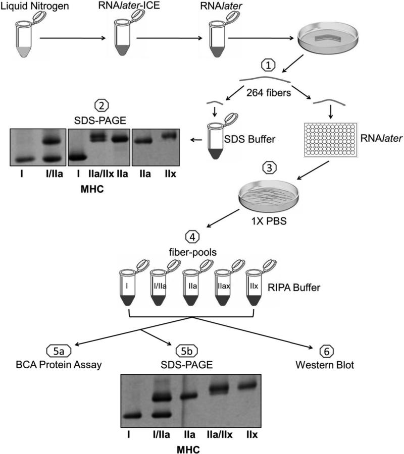
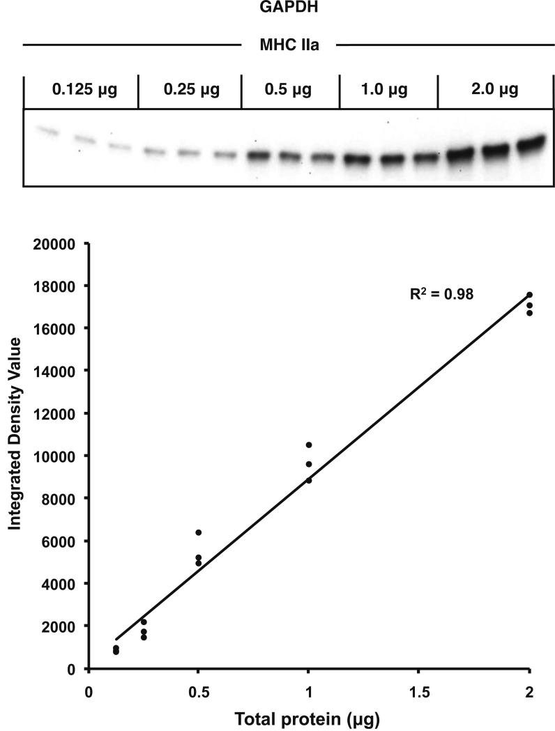
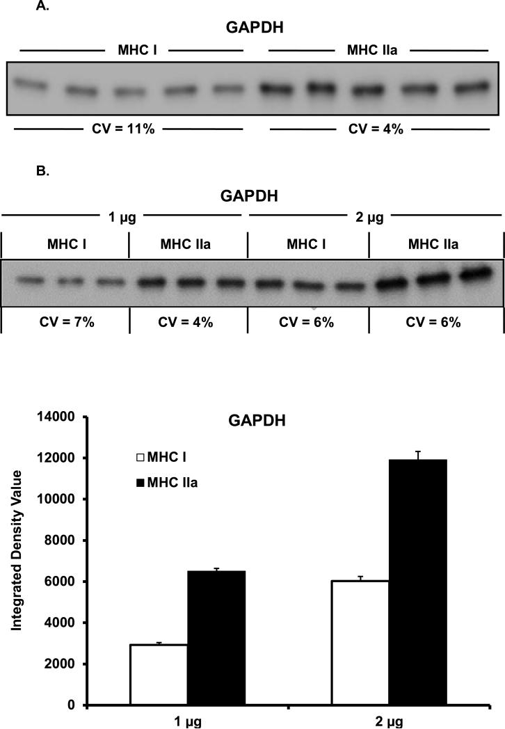
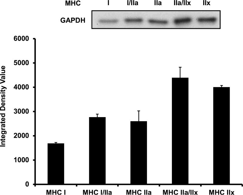
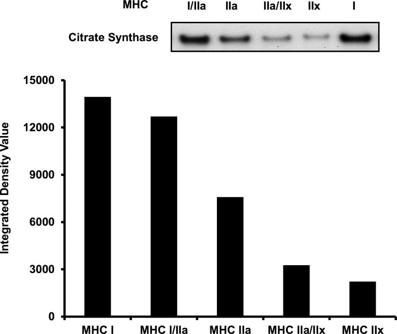
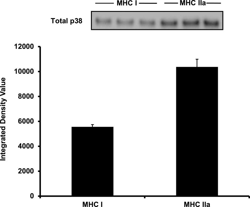
The aim of this project was to develop a method to assess fiber type specific protein content across the continuum of human skeletal muscle fibers. Individual vastus lateralis muscle fibers (n = 264) were clipped into two portions: one for sodium dodecyl sulfate-polyacrylamide gel electrophoresis (SDS-PAGE) fiber typing and one for Western blot protein identification. Following fiber type determination, fiber segments were combined into fiber type specific pools (∼20 fibers/pool) and measured for total protein quantity, glyceraldehyde 3-phosphate dehydrogenase (GAPDH), citrate synthase (CS), and total p38 content. GAPDH content was 64, 54, 160, and 138% more abundant in myosin heavy chain (MHC) I/IIa, MHC IIa, MHC IIa/IIx, and MHC IIx fibers, respectively, when compared with MHC I. Inversely, CS content was 528, 472, 242, and 47% more abundant in MHC I, MHC I/IIa, MHC IIa, and MHC IIa/IIx fibers, respectively, when compared with MHC IIx. Total p38 content was 87% greater in MHC IIa versus MHC I fibers. These data and this approach establish a reliable method for human skeletal muscle fiber type specific protein analysis. Initial results show that particular proteins exist in a hierarchal fashion throughout the continuum of human skeletal muscle fiber types, further highlighting the necessity of fiber type specific analysis.


Figures

Figure 1
Figure 1
Schematic representation of study design. 1) Two hundred and sixty four single human skeletal muscle fibers were isolated, clipped, and stored in SDS-Buffer and RNAlater. 2) All single fibers were subjected to SDS-PAGE with silver staining and classified based on migration distance. The image represents seven of these fibers. Next, single fibers of the same MHC composition were 3) briefly washed in cold 1X PBS and 4) combined into pools of 20 fibers for MHC I and MHC IIa; a pool of 11 for MHC IIx; a pool of 24 for MHC IIa/IIx and a pool of 5 for MHC IIx. 5a) The protein concentration of each fiber-pool was determined using the BCA protein assay kit. 5b) An aliquot from each fiber-pool was re-typed through SDS-PAGE to verify its MHC content. MHC = myosin heavy chain. 6) The remaining portion of each fiber-pool was utilized to assess protein content via Western blotting.
Figure 2
Figure 2
Verification of Western blot system linearity. Linearity of our system was examined by loading five different amounts of total protein (0.125, 0.25, 0.5, 1.0, and 2.0 μg) in triplicate from a MHC IIa sample, and evaluated for GAPDH content. The GAPDH signal (Integrated Density Value = IDV) was plotted against corresponding amounts of total protein.
Figure 3A & B
Figure 3A & B
Western blot GAPDH load and transfer evaluation. (A) Single MHC I and MHC IIa samples were loaded (1 μg total protein) in quintuplicate (CV = 11% and 4%, respectively). MHC IIa fibers displayed 96% more GAPDH when compared to MHC I fibers. (B) Single MHC I and MHC IIa samples were loaded in triplicate at 1 μg and 2 μg of total protein and assessed for GAPDH content. GAPDH protein content doubled when 2 μg was compared to 1 μg. MHC IIa fibers again displayed 123% and 98% more GAPDH when compared to MHC I fibers at 1 and 2 μg, respectively. MHC = myosin heavy chain. CV = coefficient of variation.
Figure 4
Figure 4
GAPDH content across the continuum of human skeletal muscle fiber types. Lanes 1-5 were loaded with 1 μg of total protein as follows: MHC I, MHC I/IIa, MHC IIa, MHC IIa/IIx, and MHC IIx. The loading scheme was repeated in lanes 6-10 (not shown) and the densitometry value for each isoform was averaged. MHC I/IIa, MHC IIa, MHC IIa/IIx, and MHC IIx displayed 64%, 54%, 160%, and 138% greater content of GAPDH when compared to MHC I, respectively. MHC = myosin heavy chain.
Figure 5
Figure 5
CS content across the continuum of human skeletal muscle fiber types. Lanes were loaded with 3 μg of total protein as follows: MHC I/IIa, MHC IIa, MHC IIa/IIx, MHC IIx, and MHC I. MHC I, MHC I/IIa, MHC IIa, and MHC IIa/IIx displayed 528%, 472%, 242%, and 47% greater content of CS when compared to MHC IIx, respectively. Due to sample limitation, only one CS fiber type continuum experiment was performed and therefore SE values are not available. MHC = myosin heavy chain. CS = Citrate Synthase.
Figure 6
Figure 6
Total p38 content in MHC I and MHC IIa fiber-pools. In 5 μg of total protein, MHC IIa fibers displayed 87% more total p38 when compared to MHC I fibers. MHC = Myosin heavy chain.
Figure 7
Figure 7
Individual human skeletal muscle fibers were studied for their contractile performance. Normalized power was 2, 6, and 12 fold greater in MHC I/IIa, MHC IIa, and MHC IIa/IIx fibers when compared to MHC I fibers. No MHC IIx fibers were studied. MHC = myosin heavy chain.

References

    1. Saltin B, Gollnick P. Handbook of Physiology. Skeletal Muscle. Am. Physiol. Soc.; Bethesda, MD: 1983. Skeletal muscle adaptability: significance for metabolism and performance.
    1. Bottinelli R, Pellegrino MA, Canepari M, Rossi R, Reggiani C. Specific contributions of various muscle fibre types to human muscle performance: an in vitro study. J Electromyogr Kinesiol. 1999;9:87–95. - PubMed
    1. Smerdu V, Karsch-Mizrachi I, Campione M, Leinwand L, Schiaffino S. Type IIx myosin heavy chain transcripts are expressed in type IIb fibers of human skeletal muscle. Am J Physiol. 1994;267:C1723–8. - PubMed
    1. Billeter R, Heizmann CW, Howald H, Jenny E. Analysis of myosin light and heavy chain types in single human skeletal muscle fibers. Eur J Biochem. 1981;116:389–95. - PubMed
    1. Gollnick PD, Armstrong RB, Saltin B, Saubert C.W.t., Sembrowich WL, Shepherd RE. Effect of training on enzyme activity and fiber composition of human skeletal muscle. J Appl Physiol. 1973;34:107–11. - PubMed

Publication types

MeSH terms

Substances