

Near-infrared molecular probes for in vivo imaging
- PMID: 22470154
- PMCID: PMC3334312
- DOI: 10.1002/0471142956.cy1227s60
Near-infrared molecular probes for in vivo imaging
Abstract
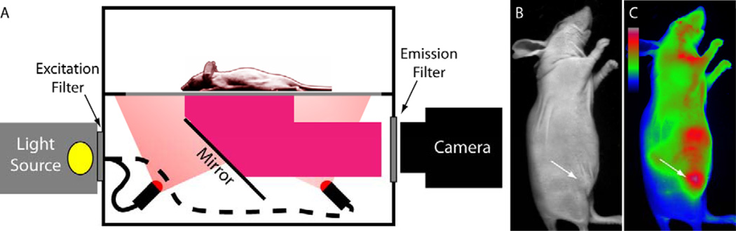
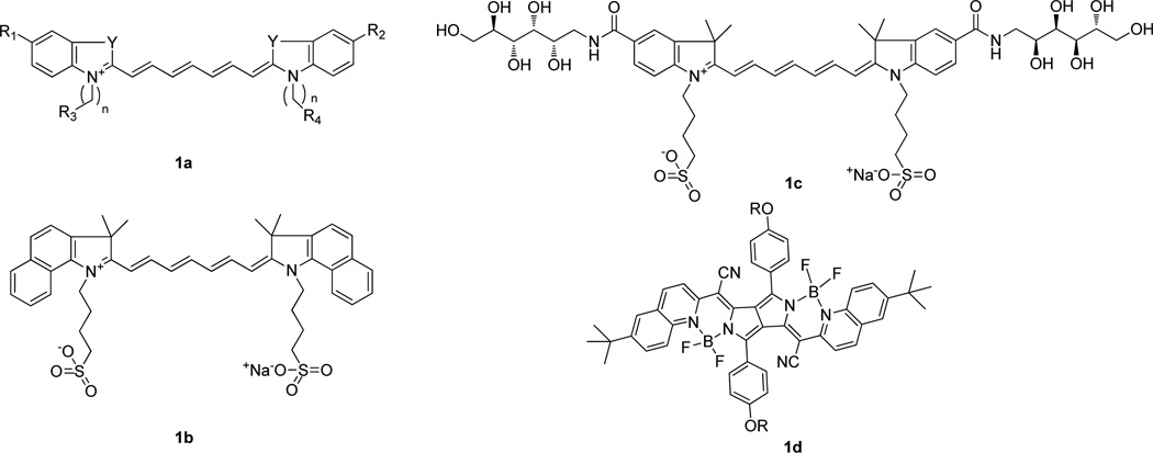
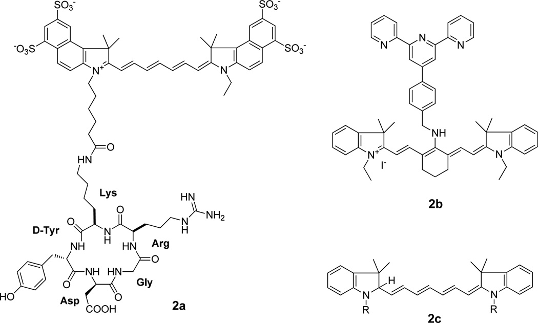
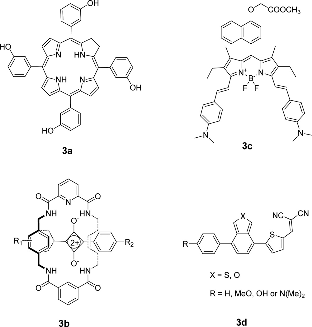
Cellular and tissue imaging in the near-infrared (NIR) wavelengths between 700 and 900 nm is advantageous for in vivo imaging because of the low absorption of biological molecules in this region. This unit presents protocols for small animal imaging using planar and fluorescence lifetime imaging techniques. Included is an overview of NIR fluorescence imaging of cells and small animals using NIR organic fluorophores, nanoparticles, and multimodal imaging probes. The development, advantages, and application of NIR fluorescent probes that have been used for in vivo imaging are also summarized. The use of NIR agents in conjunction with visible dyes and considerations in selecting imaging agents are discussed. We conclude with practical considerations for the use of these dyes in cell and small animal imaging applications.
Figures
Similar articles
-
Multifunctional materials conjugated with near-infrared fluorescent organic molecules and their targeted cancer bioimaging potentialities.Biomed Phys Eng Express. 2020 Jan 30;6(1):012003. doi: 10.1088/2057-1976/ab6e1d. Biomed Phys Eng Express. 2020. PMID: 33438589 Review.
-
A review of NIR dyes in cancer targeting and imaging.Biomaterials. 2011 Oct;32(29):7127-38. doi: 10.1016/j.biomaterials.2011.06.024. Epub 2011 Jul 2. Biomaterials. 2011. PMID: 21724249 Review.
-
Fluorescence imaging of tumors in vivo.Curr Med Chem. 2005;12(7):795-805. doi: 10.2174/0929867053507324. Curr Med Chem. 2005. PMID: 15853712 Review.
-
Long fluorescence lifetime molecular probes based on near infrared pyrrolopyrrole cyanine fluorophores for in vivo imaging.Biophys J. 2009 Nov 4;97(9):L22-4. doi: 10.1016/j.bpj.2009.08.022. Biophys J. 2009. PMID: 19883579 Free PMC article.
-
Recent Progress in Fluorescence Imaging of the Near-Infrared II Window.Chembiochem. 2018 Dec 18;19(24):2522-2541. doi: 10.1002/cbic.201800466. Epub 2018 Nov 9. Chembiochem. 2018. PMID: 30247795 Review.
Cited by
-
The Growing Development of DNA Nanostructures for Potential Healthcare-Related Applications.Adv Healthc Mater. 2019 May;8(9):e1801546. doi: 10.1002/adhm.201801546. Epub 2019 Mar 7. Adv Healthc Mater. 2019. PMID: 30843670 Free PMC article. Review.
-
Biodistribution and pharmacokinetics of Mad2 siRNA-loaded EGFR-targeted chitosan nanoparticles in cisplatin sensitive and resistant lung cancer models.Nanomedicine (Lond). 2016 Apr;11(7):767-81. doi: 10.2217/nnm.16.14. Epub 2016 Mar 16. Nanomedicine (Lond). 2016. PMID: 26980454 Free PMC article.
-
iRFP (near-infrared fluorescent protein) imaging of subcutaneous and deep tissue tumours in mice highlights differences between imaging platforms.Cancer Cell Int. 2021 May 3;21(1):247. doi: 10.1186/s12935-021-01918-8. Cancer Cell Int. 2021. PMID: 33941186 Free PMC article.
-
FLIM-FRET for Cancer Applications.Curr Mol Imaging. 2014;3(2):144-161. doi: 10.2174/2211555203666141117221111. Curr Mol Imaging. 2014. PMID: 26023359 Free PMC article.
-
M13 phage peptide ZL4 exerts its targeted binding effect on schistosoma japonicum via alkaline phosphatase.Int J Clin Exp Pathol. 2015 Feb 1;8(2):1247-58. eCollection 2015. Int J Clin Exp Pathol. 2015. PMID: 25973009 Free PMC article.
References
-
- Achilefu S, Bloch S, Markiewicz MA, Zhong TX, Ye YP, Dorshow RB, Chance B, Liang KX. Synergistic effects of light-emitting probes and peptides for targeting and monitoring integrin expression. Proceedings of the National Academy of Sciences of the United States of America. 2005;102:7976–7981. - PMC - PubMed
-
- Achilefu S, Dorshow RB, Bugaj JE, Rajagopalan R. Novel receptor-targeted fluorescent contrast agents for in vivo tumor imaging. Invest Radiol. 2000;35:479–485. - PubMed
Publication types
MeSH terms
Substances
Grants and funding
LinkOut - more resources
Full Text Sources
Other Literature Sources
Medical
Miscellaneous
