. 2012 Mar 20:3:43.
doi: 10.3389/fphar.2012.00043. eCollection 2012.

Cellular Links between Neuronal Activity and Energy Homeostasis

Affiliations

Cellular Links between Neuronal Activity and Energy Homeostasis

Pavan K Shetty et al. Front Pharmacol. .

Abstract

Neuronal activity, astrocytic responses to this activity, and energy homeostasis are linked together during baseline, conscious conditions, and short-term rapid activation (as occurs with sensory or motor function). Nervous system energy homeostasis also varies during long-term physiological conditions (i.e., development and aging) and with adaptation to pathological conditions, such as ischemia or low glucose. Neuronal activation requires increased metabolism (i.e., ATP generation) which leads initially to substrate depletion, induction of a variety of signals for enhanced astrocytic function, and increased local blood flow and substrate delivery. Energy generation (particularly in mitochondria) and use during ATP hydrolysis also lead to considerable heat generation. The local increases in blood flow noted following neuronal activation can both enhance local substrate delivery but also provides a heat sink to help cool the brain and removal of waste by-products. In this review we highlight the interactions between short-term neuronal activity and energy metabolism with an emphasis on signals and factors regulating astrocyte function and substrate supply.

Keywords: ATP; NADH; cerebral metabolism; hippocampus; lactate; neuronal metabolism; oxygen; pyruvate.


Figures
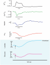
Figure 1
Figure 1
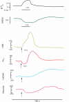
This series of traces illustrates the response to a 10–25 short stimulus train applied to the stratum radiatum of the CA1 region of an in vitro hippocampal tissue slice, and the physiological responses which can be measured in real-time during this stimulus train. These reconfigured traces have been redrawn from primary sources to be on the same time scale (shown as 120 s below). Measured responses include extracellular K+ and Ca2+ (Benninger et al., 1980) and NADH imaging and tissue PO2 (Galeffi et al., 2007). Predicted in vitro tissue slice responses are shown below with blue shading, with lactate and glucose extrapolated from in vivo measurements performed by Hu and Wilson (1997a,b). Note that extracellular K+ rises during the train and Ca2+ decreases, due to K+ release from neurons and Ca2+ uptake into neurons. NADH shows both an oxidative phase (i.e., a decrease) and a reduction phase (i.e., an increase), whereas in tissue slices the tissue oxygen can only decrease from the baseline level. Extracellular glucose can likewise only decrease, whereas lactate synthesis (stimulated by the train) results in an initial dip (due to demand) but a subsequent elevation, as lactate is further extruded from astrocytes.
Figure 2
Figure 2
This series of redrawn and rescaled traces show measured responses in vivo from intact brain (though different structures), where changes in cerebral blood flow can alter the basic responses to a stimulation train. The upper K+ and NADH traces are in response to a 25-s stimulus train at 10 Hz in the cortex (Lothman et al., 1975), showing a consistent K+ elevation and an NADH oxidation (Turner et al., 2007). The CBF and PO2 responses follow a 15-s stimulation train at 10 Hz in the cerebellum (Offenhauser et al., 2005). The glucose and lactate measurements are redrawn from Hu and Wilson (1997a,b). Note that the oxygen response shows an initial dip in the activated region then an elevation as the enhanced cerebral blood flow response occurs. There is an initial decrease and a delayed enhancement in both cerebral lactate (due to astrocytic extrusion) and glucose (due to enhanced transport into the brain) as measured extracellularly.
Figure 3
Figure 3
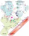
Regulation of synaptic function and cerebral blood flow: modified schematic diagram (Attwell et al., ; Kapogiannis and Mattson, 2011) showing the multiple interactions between an astrocyte, neurons, and a blood vessel. The various functions depicted show presynaptic release, glutamate receptor binding and handling, various energy transport functions between blood vessels, the astrocyte and neurons, vasoconstriction and vasodilation pathways, and extracellular molecules. Abbreviations: RYR, ryanodine receptor; ER, endoplasmic reticulum; Glu, glutamate; Gln, glutamine; IP3R, Inositol 3 phosphate receptor; Lac, lactate; Glc, glucose; Pyr, pyruvate; TCA, Tricaboxylic acid cycle; ATP, adenosine 5’- tri phosphate; Ach, acetyl choline; NE, norepinephrine; No, nitric oxide; Ad1, adenosine receptor 1; Ad2, adenosine receptor 2; mGluR, metabotropic glutamate receptor; NMDAR, N-methyl-D-aspartic acid receptor; AMPAR, 2-amino-3-(5-methyl-3-oxo-1;2- oxazol-4-yl)propanoic acid receptor; nNOS, neuronal nitric oxide synthase; eNOS, endothelial nitric oxide synthase EET, epoxyeicosatrienoic acids; AA, arachidonic acid; PGE2, prostaglandin 2; 20-HETE, 20-hydroxy arachidonic acid; PLA2, phospholipase A2; cGMP, cyclic guanosine monophosphate; P2X; purinergicreceptor P2X; P2Y, purinergic receptor P2Y; PLC, phospholipase C; AQP4, aquaporin-4; VDCC, voltage dependent calcium channel; LTCC, L-type calcium channel.

References

    1. Abbracchio M. P., Burnstock G. (1994). Purinoceptors: are there families of P2X and P2Y purinoceptors? Pharmacol. Ther. 64, 445–47510.1016/0163-7258(94)00048-4 - DOI - PubMed
    1. Ahn B. H., Kim H. S., Song S., Lee I. H., Liu J., Vassilopoulos A., Deng C. X., Finkel T. (2008). A role for the mitochondrial deacetylase Sirt3 in regulating energy homeostasis. Proc. Natl. Acad. Sci. U.S.A. 105, 14447–1445210.1073/pnas.0803790105 - DOI - PMC - PubMed
    1. Amaral A. I., Teixeira A. P., Sonnewald U., Alves P. M. (2011). Estimation of intracellular fluxes in cerebellar neurons after hypoglycemia: importance of the pyruvate recycling pathway and glutamine oxidation. J. Neurosci. Res. 89, 700–71010.1002/jnr.22571 - DOI - PubMed
    1. Amiel S. A. (1997). Hypoglycaemia in diabetes mellitus – protecting the brain. Diabetologia 40(Suppl. 2), S62–S6810.1007/s001250051404 - DOI - PubMed
    1. Anderson C. M., Swanson R. A. (2000). Astrocyte glutamate transport: review of properties, regulation, and physiological functions. Glia 32, 1–1410.1002/1098-1136(200010)32:1<1::AID-GLIA10>3.0.CO;2-W - DOI - PubMed

LinkOut - more resources