. 2012 Mar 23:3:49.
doi: 10.3389/fphar.2012.00049. eCollection 2012.

Cytoplasmic domains and voltage-dependent potassium channel gating

Affiliations

Cytoplasmic domains and voltage-dependent potassium channel gating

Francisco Barros et al. Front Pharmacol. .

Abstract

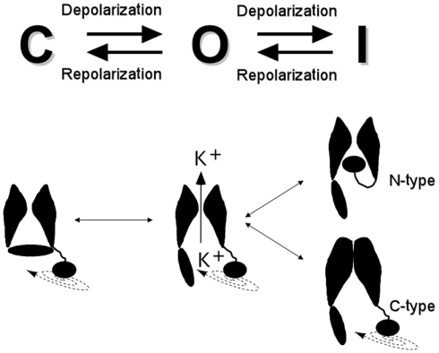
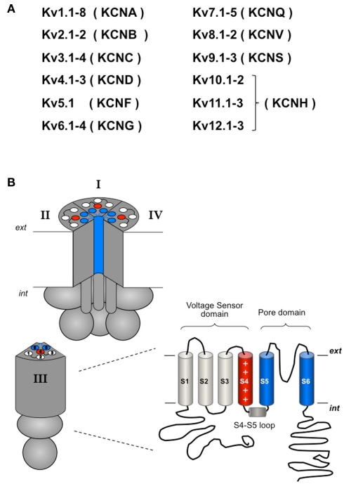
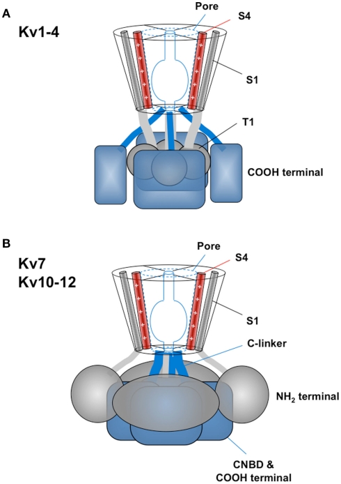
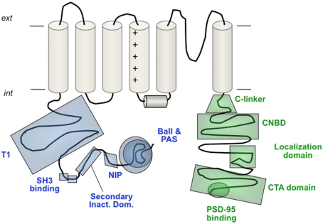
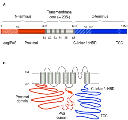
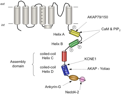
The basic architecture of the voltage-dependent K(+) channels (Kv channels) corresponds to a transmembrane protein core in which the permeation pore, the voltage-sensing components and the gating machinery (cytoplasmic facing gate and sensor-gate coupler) reside. Usually, large protein tails are attached to this core, hanging toward the inside of the cell. These cytoplasmic regions are essential for normal channel function and, due to their accessibility to the cytoplasmic environment, constitute obvious targets for cell-physiological control of channel behavior. Here we review the present knowledge about the molecular organization of these intracellular channel regions and their role in both setting and controlling Kv voltage-dependent gating properties. This includes the influence that they exert on Kv rapid/N-type inactivation and on activation/deactivation gating of Shaker-like and eag-type Kv channels. Some illustrative examples about the relevance of these cytoplasmic domains determining the possibilities for modulation of Kv channel gating by cellular components are also considered.

Keywords: activation/deactivation gating; cytoplasmic domains; inactivation gating; potassium channel; structure–function relationships; voltage-dependent gating.


Figures

Figure 1
Figure 1
Schematic cartoon of the conformational states and voltage-driven gating modifications of Kv channels. The drawings at the bottom represent two α-subunits and a symbolic cytoplasmic gate, with one diffusible ball-and-chain structure attached to their cytoplasmic face. N-type inactivation as a consequence of plugging of the pore after opening the cytoplasmic activation gate, and C-type inactivation by collapsing of the selectivity filter gate, are represented on the right.
Figure 2
Figure 2
(A) Overview of the Kv channels subfamilies. (B) Schematic representation of the tetrameric organization of a Kv channel. A structural folding model of one of the four α subunits is shown on the right. Note that in contrast to the considerable homogeneity in the transmembranal core and the loops linking the transmembrane helices, large variations in the relative size and positioning of the amino and carboxy terminals are found in the different Kv channels. For more explanations, see text.
Figure 3
Figure 3
Basic patterns of Kv channel cytoplasmic domains structural organization. (A) General organization of the Kv1–Kv4 channels group with the T1 “tetramerization domains” hanging centrally below the transmembranal core and attached to it through four linkers continued from the first transmembrane helices. In this case the C-terminal structures probably track to the periphery surrounding T1 and extending to its bottom. (B) General cytoplasmic architecture of the Kv7 and Kv10–Kv12 channels characterized by a carboxy terminus (i.e., the C-linker/CNBD region of the Kv10–Kv12 channels or the A–D helical regions of the Kv7 channels) forming a compact tetrameric structure in a central position immediately below the cytoplasmic pore opening. In this case the amino terminus probably surrounds the C-terminus and extends to its bottom establishing extensive contacts with its top and side surfaces. Note that in both models the initial amino terminal structures (ball-like and eag/PAS domains for Kv1–Kv4 and Kv10–Kv12, respectively) are likely to interact with the gate surroundings in the transmembrane core. S4 segment is also depicted as a reference. See text for details.
Figure 4
Figure 4
Schematic view of different structural and/or functional domains recognized at the cytoplasmic ends of Kv channels. In the amino terminus these include the ball-like structure responsible for fast N-type inactivation, the eag/PAS domain of the eag-like channels, the NIP domain that protects Kv1.6 channels against rapid inactivation, the secondary inactivation domain reported for Kv1.4 channels, the double SH3 binding domain of Kv1.5, and the T1 tetramerization domain (also called NAB) of the Kv1–Kv4 channels. In the carboxy terminus the domains shown correspond to the C-linker and cNBD encountered in the eag-like channels, the localization domain and the C-terminal activation (CTA) domain of Kv2.1 and the post-synaptic density protein (PSD-95)-binding domain of some Kv1 channels. A more detailed view of the Kv7 channels carboxy terminus organization is shown schematically in Figure 6. For more explanations, see text. Note that not all the depicted domains pertain to the same Kv channel.
Figure 5
Figure 5
Structural organization of the hERG K+ channel. (A) Schematic linear diagram of the hERG channel protein. The regions corresponding to the eag/PAS (residues 1–135) and the proximal domains up to the first transmembrane helix are shown as striped and solid red bars, respectively. The transmembranal core region containing the six transmembrane helices and the carboxy terminus are shown colored in gray and blue, respectively. TCC indicates the proposed location of a tetramerization coiled-coil at the C-terminus. The size of every domain is represented on a horizontal scale proportional to the total length of the protein. (B) Schematic representation of a hERG channel α-subunit showing the proposed relative positioning of the PAS region in the amino terminus (dotted line), and the flexible tail of the amino end (initial solid red line segment) and the amphipathic α-helix separating them (red rectangle) pointing toward the S4–S5 linker on the cytoplasmic surface of the channel core. The proximity between the initial regions of the N-terminus and the C-linker/cNBD domains that are directly linked to the cytoplasmic gate at the bottom of helix S6 is also used to illustrate the possible existence of additional interactions between these channel structures. For more explanations, see text.
Figure 6
Figure 6
Schematic representation of the carboxy-terminal tail structures and/or interaction sites present in Kv7 channels. The four helical regions (helices A–D) conserved in all family members are shown as cylinders. Formation of coiled-coil assemblies at the level of helices C and D is indicated. The proposed location of the conserved interaction sites with calmodulin (CaM) and phosphatidylinositol 4,5-bisphosphate (PIP2), with A-kinase anchoring protein 79/150 (AKAP79/150) in Kv7.2, with the auxiliary β subunit KCNE1 in Kv7.1, with AKAP-Yotiao in Kv7.1, with ankyrin-G in Kv7.2–Kv7.3 and with ubiquitin-protein ligase Nedd4-2 in Kv7.1 and Kv7.2–Kv7.3, are also shown. Phosphorylation sites by Src kinase in helix A and by protein kinase C in helix B are indicated by an encircled P.

References

    1. Adair B., Nunn R., Lewis S., Dukes I., Philipson L., Yeager M. (2008). Single particle image reconstruction of the human recombinant Kv2.1 channel. Biophys. J. 94, 2106–211410.1529/biophysj.107.118562 - DOI - PMC - PubMed
    1. Adams J. P., Anderson A. E., Varga A. W., Dineley K. T., Cook R. G., Pfaffinger P. J., Sweatt J. D. (2000). The A-type potassium channel Kv4.2 is a substrate for the mitogen-activated protein kinase ERK. J. Neurochem. 75, 2277–228710.1046/j.1471-4159.2000.0752277.x - DOI - PubMed
    1. Alaimo A., Gómez-Posada J. C., Aivar P., Etxeberría A., Rodriguez-Alfaro J. A., Areso P., Villarroel A. (2009). Calmodulin activation limits the rate of KCNQ2 K+ channel exit from the endoplasmic reticulum. J. Biol. Chem. 284, 20668–2067510.1074/jbc.M109.019539 - DOI - PMC - PubMed
    1. Alonso-Ron C., Barros F., Manso D. G., Gómez-Varela D., Miranda P., Carretero L., Domínguez P., de la Peña P. (2009). Participation of HERG channel cytoplasmic structures on regulation by the G protein-coupled TRH receptor. Pflügers Arch. 457, 1237–125210.1007/s00424-008-0599-x - DOI - PubMed
    1. Alonso-Ron C., de la Peña P., Miranda P., Domínguez P., Barros F. (2008). Thermodynamic and kinetic properties of amino-terminal and S4–S5 loop HERG channel mutants under steady-state conditions. Biophys. J. 94, 3893–391110.1529/biophysj.107.116731 - DOI - PMC - PubMed

LinkOut - more resources