. 2012;7(3):e33625.
doi: 10.1371/journal.pone.0033625. Epub 2012 Mar 28.

Early developing pig embryos mediate their own environment in the maternal tract

Affiliations

Early developing pig embryos mediate their own environment in the maternal tract

Carmen Almiñana et al. PLoS One. 2012.

Abstract

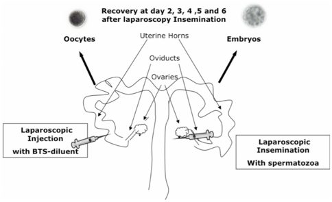
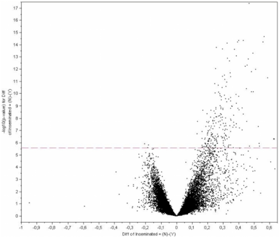
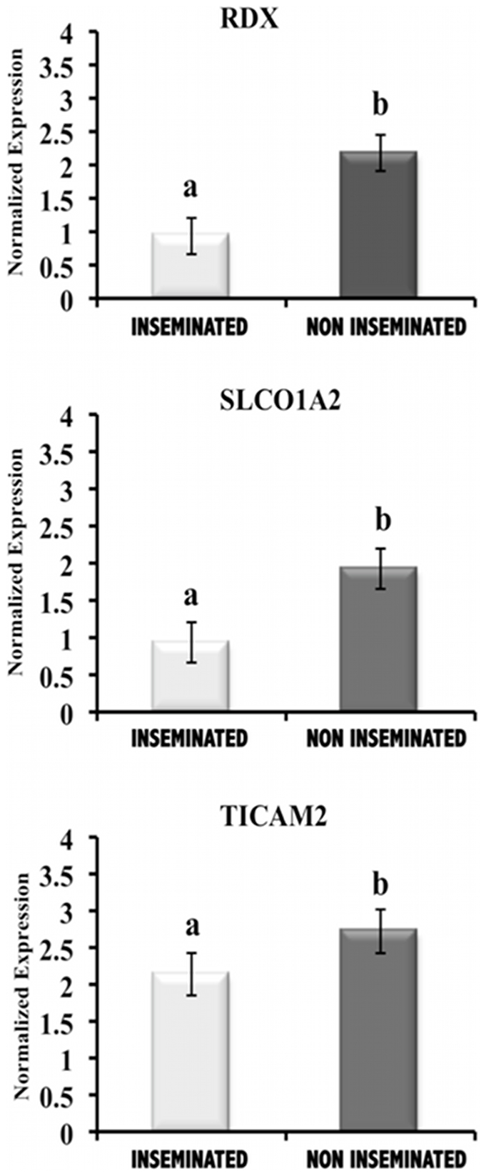
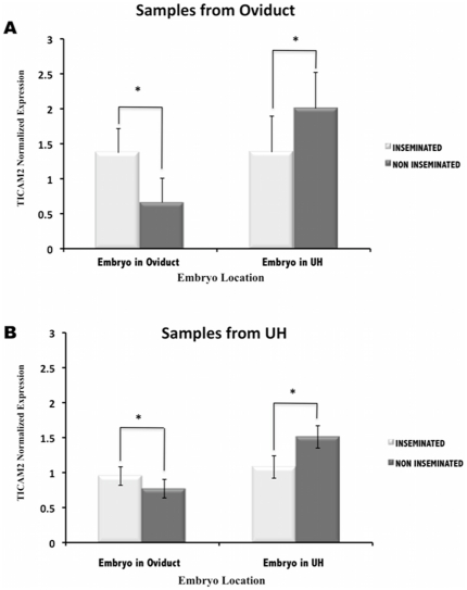
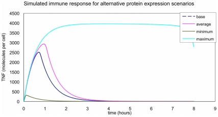
The maternal tract plays a critical role in the success of early embryonic development providing an optimal environment for establishment and maintenance of pregnancy. Preparation of this environment requires an intimate dialogue between the embryo and her mother. However, many intriguing aspects remain unknown in this unique communication system. To advance our understanding of the process by which a blastocyst is accepted by the endometrium and better address the clinical challenges of infertility and pregnancy failure, it is imperative to decipher this complex molecular dialogue. The objective of the present work is to define the local response of the maternal tract towards the embryo during the earliest stages of pregnancy. We used a novel in vivo experimental model that eliminated genetic variability and individual differences, followed by Affymetrix microarray to identify the signals involved in this embryo-maternal dialogue. Using laparoscopic insemination one oviduct of a sow was inseminated with spermatozoa and the contralateral oviduct was injected with diluent. This model allowed us to obtain samples from the oviduct and the tip of the uterine horn containing either embryos or oocytes from the same sow. Microarray analysis showed that most of the transcripts differentially expressed were down-regulated in the uterine horn in response to blastocysts when compared to oocytes. Many of the transcripts altered in response to the embryo in the uterine horn were related to the immune system. We used an in silico mathematical model to demonstrate the role of the embryo as a modulator of the immune system. This model revealed that relatively modest changes induced by the presence of the embryo could modulate the maternal immune response. These findings suggested that the presence of the embryo might regulate the immune system in the maternal tract to allow the refractory uterus to tolerate the embryo and support its development.


Conflict of interest statement

Competing Interests: The authors have declared that no competing interests exist.

Figures

Figure 1
Figure 1. Schematic representation of the experimental design.
Sows were subjected to laparoscopic surgery. While one oviduct was subjected to laparoscopic insemination with (3×10⁵ spermatozoa/100 µl) spermatozoa (inseminated side), the contralateral oviduct was injected with BTS-diluent containing no sperm (non inseminated side). Oviductal and uterine horn samples as well as flushings were collected from both horns at day 2, 3, 4, 5 and 6 after laparoscopy insemination by hysterectomy. The presence of embryos at different stages of pregnancy in one horn (inseminated side), and the existence of unfertilized oocytes in the other horn (non inseminated side) were verified by careful examination of flushings under a stereomicroscope.
Figure 2
Figure 2. Schematic diagram of the components of TLR4 pathway model.
This figure shows a schema of the components and interaction of the published TLR4 computational model from An and Faeder model .
Figure 3
Figure 3. Mapping of differentially expressed genes onto Volcano plot.
A Volcano plot depicting significant changes in gene expression between uterine horn in the presence of the blastocyst (Inseminated side) and uterine horn in the presence of oocytes (Non inseminated side). Each of the 23,124-oligonucleotide probes is plotted and probes showing significant differences in gene expression (210 probes, p<0.05) are above the red broken line.
Figure 4
Figure 4. Validation of the microarray results by qPCR analysis.
RDX (radixin), SLCO1A2 (solute carrier organic anion transporter family member 1A2) and TICAM2 (TIR domain- containing adapter molecule 2) expression values (normalized based on ß–actin expression values) in uterine horn samples in the presence of embryos at the blastocyst stage (Inseminated) compared to the presence of oocytes (Non Inseminated). The expression of all transcripts in the uterine horn in the presence of blastocyst was significantly different from that of oocyte in the uterine horn (P<0.05).
Figure 5
Figure 5. Transcripts differentially expressed in uterine horn in presence of embryos organized into functional categories.
The proportions of the differentially expressed transcripts were organized into 4 major categories (first level) and different subcategories (39) (second level) on the basis KEGG PATHWAY database hierarchy The central pie chart represents the first level of organization and the lateral pies display the second level of organization: a total of 39 subcategories containing the 84 different pathways associated with the differentially expressed transcripts.
Figure 6
Figure 6. Spatial–course of TICAM2 expression in oviduct and uterine horn (UH) samples.
TICAM2 expression values (normalized based on ß–actin expression values) in oviduct (A) and UH samples (B) in the presence of embryos at different stages of development (Inseminated) and in the presence of oocytes (Non inseminated). Embryo was located in oviduct at 2 cells and 4 cells embryo stage; Embryo was located in uterine horn at 4 cells, morula and blastocyst). *(P<0.05).
Figure 7
Figure 7. Time–course of TICAM2 expression in oviduct and uterine horn (UH) samples.
TICAM2 expression values (normalized based on ß–actin expression values) in oviduct (A) and UH samples (B) in the presence of embryos at different stages of development (Inseminated) and in the presence of oocytes (Non inseminated). *(P<0.05).
Figure 8
Figure 8. TNF profiles for the different scenarios.
The figure shows the different response of the model in terms of produced TNF in the presence of oocytes (base) and embryos (maximum, average and minimum response).

References

    1. Haouzi D, Assou S, Mahmoud K, Tondeur S, Reme T, et al. Gene expression profile of human endometrial receptivity: comparison between natural and stimulated cycles for the same patients. Hum Reprod. 2009;24:1436–1445. - PMC - PubMed
    1. Fazeli A. Maternal communication with gametes and embryos. Theriogenology. 2008;70:1182–1187. - PubMed
    1. Pope W. Embryonic mortality in swine; In: Geisert Rd ZM, editor. Boca Raton: CRS Press; 1994. pp. 53–77.
    1. Simon C, Moreno C, Remohi J, Pellicer A. Molecular interactions between embryo and uterus in the adhesion phase of human implantation. Hum Reprod. 1998;13(Suppl 3):219–232; discussion 233–216. - PubMed
    1. Wolf E, Arnold GJ, Bauersachs S, Beier HM, Blum H, et al. Embryo-maternal communication in bovine - strategies for deciphering a complex cross-talk. Reprod Domest Anim. 2003;38:276–289. - PubMed

Publication types

Substances