. 2012;7(3):e33693.
doi: 10.1371/journal.pone.0033693. Epub 2012 Mar 21.

Methylphenidate exposure induces dopamine neuron loss and activation of microglia in the basal ganglia of mice

Affiliations

Methylphenidate exposure induces dopamine neuron loss and activation of microglia in the basal ganglia of mice

Shankar Sadasivan et al. PLoS One. 2012.

Erratum in

  • PLoS One. 2012;7(5): doi/10.1371/annotation/c76da2c1-ccb8-4797-94c1-359d3ceceeda

Abstract

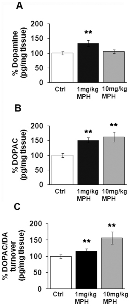
Background: Methylphenidate (MPH) is a psychostimulant that exerts its pharmacological effects via preferential blockade of the dopamine transporter (DAT) and the norepinephrine transporter (NET), resulting in increased monoamine levels in the synapse. Clinically, methylphenidate is prescribed for the symptomatic treatment of ADHD and narcolepsy; although lately, there has been an increased incidence of its use in individuals not meeting the criteria for these disorders. MPH has also been misused as a "cognitive enhancer" and as an alternative to other psychostimulants. Here, we investigate whether chronic or acute administration of MPH in mice at either 1 mg/kg or 10 mg/kg, affects cell number and gene expression in the basal ganglia.

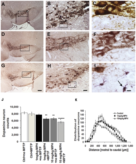
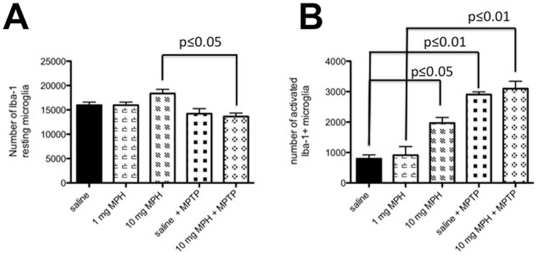
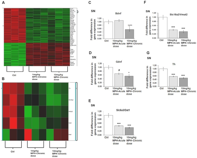
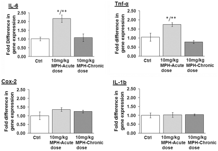
Methodology/principal findings: Through the use of stereological counting methods, we observed a significant reduction (∼20%) in dopamine neuron numbers in the substantia nigra pars compacta (SNpc) following chronic administration of 10 mg/kg MPH. This dosage of MPH also induced a significant increase in the number of activated microglia in the SNpc. Additionally, exposure to either 1 mg/kg or 10 mg/kg MPH increased the sensitivity of SNpc dopaminergic neurons to the parkinsonian agent 1-methyl-4-phenyl-1,2,3,6-tetrahydropyridine (MPTP). Unbiased gene screening employing Affymetrix GeneChip® HT MG-430 PM revealed changes in 115 and 54 genes in the substantia nigra (SN) of mice exposed to 1 mg/kg and 10 mg/kg MPH doses, respectively. Decreases in the mRNA levels of gdnf, dat1, vmat2, and th in the substantia nigra (SN) were observed with both acute and chronic dosing of 10 mg/kg MPH. We also found an increase in mRNA levels of the pro-inflammatory genes il-6 and tnf-α in the striatum, although these were seen only at an acute dose of 10 mg/kg and not following chronic dosing.

Conclusion: Collectively, our results suggest that chronic MPH usage in mice at doses spanning the therapeutic range in humans, especially at prolonged higher doses, has long-term neurodegenerative consequences.


Conflict of interest statement

Competing Interests: The authors have declared that no competing interests exist.

Figures

Figure 1
Figure 1. Chronic MPH administration affects SNpc dopamine neuron numbers and induces microgliosis.
Representative images of the substantia nigra pars compacta (SNpc) from brains of animals treated with either saline (A–C), 1 mg/kg MPH (D–F) or 10 mg/kg MPH(G–I). The images presented are at 4×, 20× and 100×, respectively. The brain sections have been immunostained with anti-TH (brown) to identify dopaminergic neurons and anti-Iba-1 (purple) to identify microglia. (J) Stereological estimates of dopamine neuron number in substantia nigra pars compacta (SNpc) in animals administered saline (ctrl), saline+MPTP (ctrl+MPTP), 1 mg/kg MPH, 1 mg/kg MPH+MPTP, 10 mg/kg MPH and 10 mg/kg MPH+MPTP. Saline, 1 mg/kg MPH and 10 mg/kg were administered for 90 days following a one-week drug washout period before 4×20 mg/kg MPTP was injected (n = 10). (K) The distribution of dopamine neurons along the rostral-caudal axis in SNpc following chronic administration (90 days) of saline (control), 1 mg/kg MPH and 10 mg/kg MPH. **p≤0.01 compared to saline-treated controls; ***p≤0.001 10 mg/kg MPH compared to saline-control (ctrl), control+MPTP (ctrl+MPTP) and 1 mg/kg MPH+ no MPTP (n = 10). One-way ANOVA statistical test was performed to draw comparisons between the different groups followed by Bonferroni post-hoc tests. Scale bars (A,D,G), 200 µms; (B,E,H), 40 µms and (C,F,I), 8 µms.
Figure 2
Figure 2. Chronic exposure to high dose MPH results in microglial cell activation in the SNpc.
Stereological estimates of Iba-1 positive microglia cells in the SNpc (A) the total number of morphologically-resting microglia and (B) the total number of morphologically-activated microglia following chronic administration of either saline (ctrl), 1 mg/kg MPH, 10 mg/kg MPH and 10 mg/kg MPH+MPTP. (n = 5). One-way ANOVA statistical test was performed to draw comparisons between the different groups followed by Bonferroni post-hoc tests.
Figure 3
Figure 3. Chronic MPH dosing alters dopamine turnover in the striatum.
Striata were microdissected from the brains of mice administered 90 days of saline (Ctrl), 1 mg/kg MPH or 10 mg/kg MPH and were processed for HPLC analyses. Total striatal levels of (A) dopamine and (B) the dopamine metabolite 3,4-dihydroxyphenylacetic acid (DOPAC) are presented as percentage of saline-treated controls (Ctrl). (C) Dopamine turnover is presented as the ratio of DOPAC/DA. *p≤0.01 compared to saline-controls (Ctrl) (n = 8). One-way ANOVA statistical test was performed to draw comparisons between the different groups followed by Bonferroni post-hoc tests.
Figure 4
Figure 4. Acute and chronic administration of MPH alters gene expression in the substantia nigra (SN).
(A) Heat map representation of gene expression changes following chronic administration of either 1 mg/kg MPH or 10 mg/kg MPH in the SN (n = 3). qPCR analysis demonstrating normalized fold-change expression of (B) bdnf, (C) gdnf, (D) dat1(slc6a3), (E) vmat2(slc18a2) and (F) th mRNA in SN (n = 3). *p≤0.02 vs saline-controls (ctrl); **p≤0.02 vs saline-controls and 10 mg/kg MPH-acute dose; #p≤0.02 10 mg/kg MPH acute-dose vs saline-controls (ctrl). One-way ANOVA statistical test was performed to draw comparisons between the different groups followed by Bonferroni post-hoc tests.
Figure 5
Figure 5. mRNA expression of pro-inflammatory genes following acute administration of 10 mg/kg MPH in the striatum.
Fold change values in mRNA expression presented are normalized against saline controls, in the striatum. The genes probed for include (A) il-6, (B) tnf-á, (C) cox2 and (D) il-1b. *p≤0.02 compared to saline-controls (ctrl); **p≤0.02 compared to 10 mg/kg MPH-chronic dose, (n = 3). One-way ANOVA statistical test was performed to draw comparisons between the different groups followed by Bonferroni post-hoc tests.

References

    1. Arnsten AF. Stimulants: Therapeutic actions in ADHD. Neuropsychopharmacology. 2006;31:2376–2383. - PubMed
    1. Capp PK, Pearl PL, Conlon C. Methylphenidate HCl: therapy for attention deficit hyperactivity disorder. Expert Rev Neurother. 2005;5:325–331. - PubMed
    1. Hirai N, Nishino S. Recent advances in the treatment of narcolepsy. Curr Treat Options Neurol. 2011;13:437–457. - PubMed
    1. Volkow ND, Wang GJ, Fowler JS, Fischman M, Foltin R, et al. Methylphenidate and cocaine have a similar in vivo potency to block dopamine transporters in the human brain. Life Sci. 1999;65:L7–12. - PubMed
    1. Johnston LD, O'Malley PM, Bachman JG, Schulenberg JE. Monitoring the Future national survey results on drug use, 1975–2010. 2011. pp. 1–312. Volume II: College students and adults ages 19–50. The University of Michigan.

Publication types

MeSH terms

Substances