. 2009;3(6):21-5.
doi: 10.3941/jrcr.v3i6.218. Epub 2009 Jun 1.

Significance of reversal of diastolic blood flow in the evolution of testicular infarction as a complication of epididymo-orchitis

Affiliations

Significance of reversal of diastolic blood flow in the evolution of testicular infarction as a complication of epididymo-orchitis

Robert Marks et al. J Radiol Case Rep. 2009.

Abstract

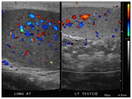
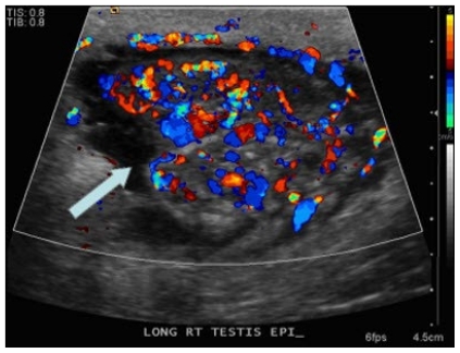
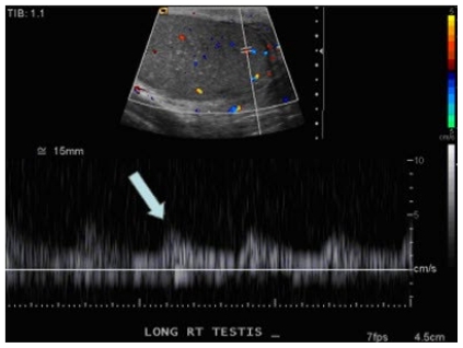
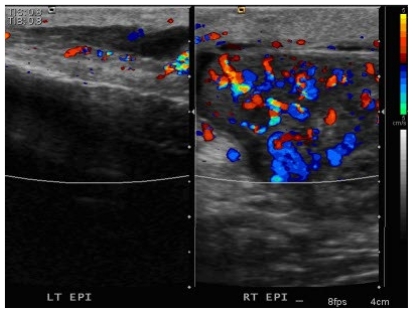
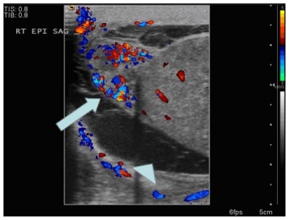
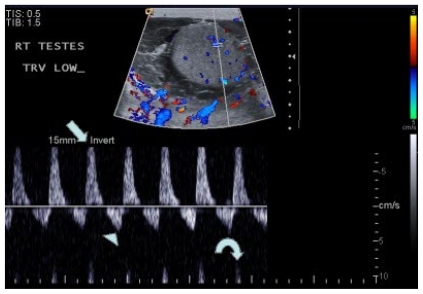
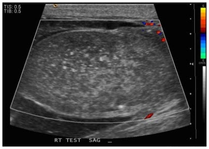
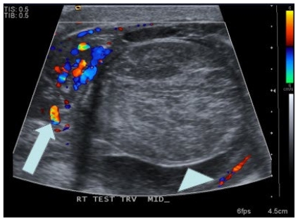
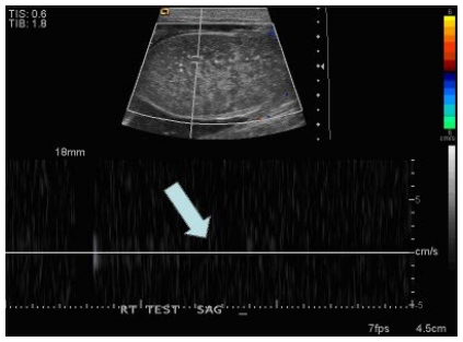
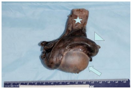
We report a case of a 50-year-old male who presented to the Emergency Department and was diagnosed with epididymo-orchitis. Sonographic evaluation of the testicle initially showed a normal, low resistance color Doppler waveform. The patient was admitted to the hospital. A follow up sonogram two days later demonstrated reversal of diastolic arterial flow on Pulse-Wave color Doppler imaging. Reversal of diastolic blood flow in testicular color Doppler sonography is a sign of impending infarction. On hospital day 6, the patient had a follow up ultrasound which demonstrated infarction of the testicle. Pathology confirmed the diagnosis and the tissue culture grew E. coli and Candida Albicans. This case documents the rapid progression of epididymo-orchitis with a normal spectral waveform to testicular infarction with reversal of diastolic blood flow on color Doppler imaging as a sign of impending infarction.

Keywords: Testicular infarction; epididymo-orchitis; reversal of diastolic flow.


Figures

Figure 1
Figure 1
50-year-old male with right sided epididymo-orchitis. Side by side longitudinal ultrasound image on day of admission demonstrates an enlarged right testicle (measuring 5.0 cm × 3.1 cm × 2.8 cm) that is slightly hypoechoic when compared to the left testicle (measuring 4.0 cm × 2.5 cm × 2.0 cm). The right testicle also demonstrates subtle hyperemia compared to the left. These findings are consistent with right sided orchitis.
Figure 2
Figure 2
50-year-old male with right sided epididymo-orchitis. Sonogram of the right epididymis on day of admission (arrow) demonstrates an enlarged right epididymis (measuring 1.4 cm × 1.6 cm).
Figure 3
Figure 3
50-year-old male with right sided epididymo-orchitis. Color Doppler sonogram of the right epididymis on day of admission (arrow) demonstrates increased vascularity throughout the epididymis consistent with epididymitis.
Figure 4
Figure 4
50-year-old male with right sided epididymo-orchitis. Spectral waveform on color Doppler imaging on day of admission (arrow) demonstrates a normal, low resistance arterial waveform in the right testicle.
Figure 5
Figure 5
50-year-old male with right sided epididymo-orchitis. Side by side color Doppler image of the right and left epididymis on day of admission demonstrates an enlarged and hypervascular right epididymis (measuring 1.4 cm × 1.6 cm) compared to the left (measuring 1.1 cm × 0.7 cm) consistent with right sided epididymitis.
Figure 6
Figure 6
50-year-old with right sided epididymo-orchitis. Color Doppler sonogram of the right testicle and epididymis 2 days after admission demonstrates an enlarged (measuring 1.6 cm × 1.0 cm), hyperemic right epididymis (arrow) consistent with epididymitis. There has been development of a complex right hydrocele (arrowhead) without internal blood flow representing a pyocele.
Figure 7
Figure 7
50-year-old with right sided epididymo-orchitis. Spectral waveform on color Doppler imaging 2 days after admission demonstrates reversal of diastolic blood flow. The peak systolic flow is elevated (arrow), and the peak diastolic flow is reversed (arrowhead). Incidentally noted is aliasing artifact (curved arrow). In the setting of infection, this type of waveform is concerning for impending infarction. Also demonstrated is the previously described complex right hydrocele, representing a pyocele.
Figure 8
Figure 8
50-year-old male with right sided epididymo-orchitis. Color Doppler sonogram of the right testicle 6 days after admission demonstrates no appreciable color flow within the right testicle which appears more heterogeneous in echotexture. These findings are consistent with testicular infarction.
Figure 9
Figure 9
50-year-old male with right sided epididymo-orchitis. Color Doppler image of the right epididymis (arrow) and right testicle (arrowhead) 6 days after admission demonstrate continued hyperemia of the right epididymis with no appreciable flow in the right testicle, consistent with infarction. Again noted is the complex right hydrocele, representing a pyocele.
Figure 10
Figure 10
50-year-old male with right sided epididymo-orchitis. Spectral waveform image on color Doppler imaging 6 days after admission demonstrates no appreciable spectral waveform (arrow) in the right testicle confirming testicular infarction.
Figure 11
Figure 11
50-year-old male with right sided epididymo-orchitis that lead to testicular infarction. Gross surgical specimen demonstrates a gray-brown appearance to the right testicle (arrow), epididymis (arrowhead), and spermatic cord (star). These findings are consistent with acute and chronic epididymo-orchitis with hemorrhage and necrosis.
Figure 12
Figure 12
50-year-old male with right sided epididymo-orchitis that lead to testicular infarction. Gross surgical specimen demonstrates a tan-white to gray-brown appearance to the right testicle and epididymis. Noted in the distal portion of the right testicle are multiple purulent nodules measuring up to 0.3cm. These findings are consistent with acute and chronic epididymo-orchitis with hemorrhage and necrosis.
Figure 13
Figure 13
50-year-old male with right sided epididymo-orchitis that lead to testicular infarction. Gross bivalved surgical specimen demonstrates a tan-red appearance to the right testicle (arrow) consistent with hemorrhage with a thin rim of normal apparent normal testicular parenchyma remaining. The epididymis (arrowhead) has surface hemorrhage and fibrosis throughout. The spermatic cord (star) is tan-white consistent with fibrosis with streak hemorrhages running throughout. These findings are consistent with acute and chronic epididymo-orchitis with hemorrhage and necrosis.

References

    1. Sue SR, Pelucio M, Gibbs M. Testicular infarction in a patient with epididymitis. Acad Emerg Med. 1998 Nov;5(11):1128–30. - PubMed
    1. Bude RO, Rubin JM. Relationship between the resistive index and vascular compliance and resistance. Radiology. 1999 May;211(2):411–7. - PubMed
    1. Mihmanli I, Kantarci F, Kulaksizoglu H, et al. Testicular size and vascular resistance before and after hydrocelectomy. AJR Am J Roentgenol. 2004 Nov;183(5):1379–85. - PubMed
    1. Sanders LM, Haber S, Dembner A, Aquino A. Significance of reversal of diastolic flow in the acute scrotum. J Ultrasound Med. 1994 Feb;13(2):137–9. - PubMed
    1. Lockhart ME, Wells CG, Morgan DE, Fineberg NS, Robbin ML. Reversed diastolic flow in the renal transplant: perioperative implications versus transplants older than 1 month. AJR Am J Roentgenol. 2008 Mar;190(3):650–5. - PubMed

LinkOut - more resources