

doi: 10.3941/jrcr.v3i7.269.
Epub 2009 Jul 1.
Median palatine cyst
Affiliations
- PMID: 22470670
- PMCID: PMC3303325
- DOI: 10.3941/jrcr.v3i7.269
Item in Clipboard
Median palatine cyst
J Radiol Case Rep.
2009.
Abstract
Median palatine cysts are rare, non-odontogenic fissural cysts of the hard palate. These cysts occur in the midline of the hard palate, behind the incisive canal. Only two case reports have documented these cysts on multi-detector computed tomography (MDCT), neither giving detailed descriptions of the cysts. Knowledge of their existence is important and should not be confused with malignant tumors. We present the first case describing the MDCT characteristics of the median palatine cyst.
Keywords: Computed Tomography; Fissural cyst; Median palatine cyst; Nasopalatine cyst.
Figures

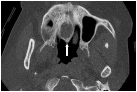
Contrast enhanced axial MDCT image in bone windows at the level of the maxilla. The median palatine cyst appears as a well circumscribed, expansile, ovoid cyst located in the midline of the hard palate (arrow).

Contrast enhanced MDCT coronal reformatted image in bone windows through the paranasal sinuses. There is a well circumscribed, expansile, ovoid cyst located in the midline of the hard palate (arrow). Note the elevation of the floor of the nasal cavity and extension into the left nares.

Contrast enhanced MDCT sagittal reformatted image in bone windows through the paranasal sinuses. Again noted is a well circumscribed, expansile, ovoid cyst located in the hard palate. There is no communication of the median palatine cyst (arrowhead) with the incisive canal (arrow).
Noncontrast axial MDCT image in bone windows at the level of the maxilla. Follow up imaging 10 months later demonstrated stable size and appearance of the median palatine cyst (arrow).
References
-
- Karacal N, Ambarcoglu O, Kutlu N. Median palatine cyst: Report of an unusual entity. Plast Reconstr Surg. 2005 Apr;115(4):1213–4. - PubMed
-
- Hadi U, Younes A, Ghosseini S, Tawil A. Median palatine cyst: An unusual presentation of a rare entity. Br J Oral Maxillofac Surg. 2001 Aug;39(4):278–81. - PubMed
-
- Gingell JC, Levy BA, Depaola LG. Median palatine cysts. J Oral Maxillofac Surg. 1985 Jan;43(1):47–51. - PubMed
-
- Zachariades N, Papanikolaou S. The median palatal cyst: does it exist? Report of three cases with oro-medical implications. J Oral Med. 1984 Jul-Sep;39(3):173–6. - PubMed
-
- Gordon NC, Swann NP, Hansen LS. Median palatine cyst and maxillary antral osteoma. J Oral Surg. 1980 May;38(5):361–5. - PubMed
LinkOut - more resources
Full Text Sources
Other Literature Sources