. 2010;4(11):1-8.
doi: 10.3941/jrcr.v4i11.462. Epub 2010 Nov 1.

CT and MR findings in extramedullary haematopoiesis with biliary system encasement: a case report

Affiliations

CT and MR findings in extramedullary haematopoiesis with biliary system encasement: a case report

Alfredo La Fianza et al. J Radiol Case Rep. 2010.

Abstract

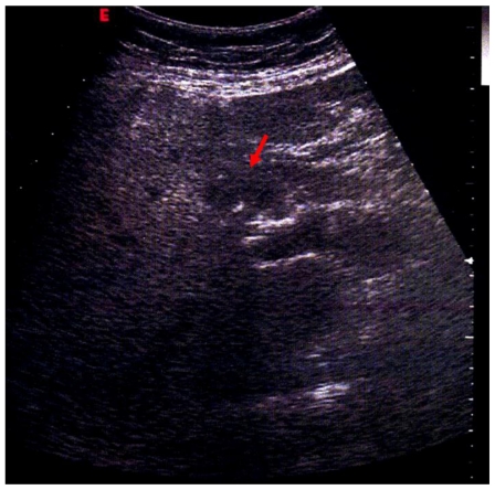
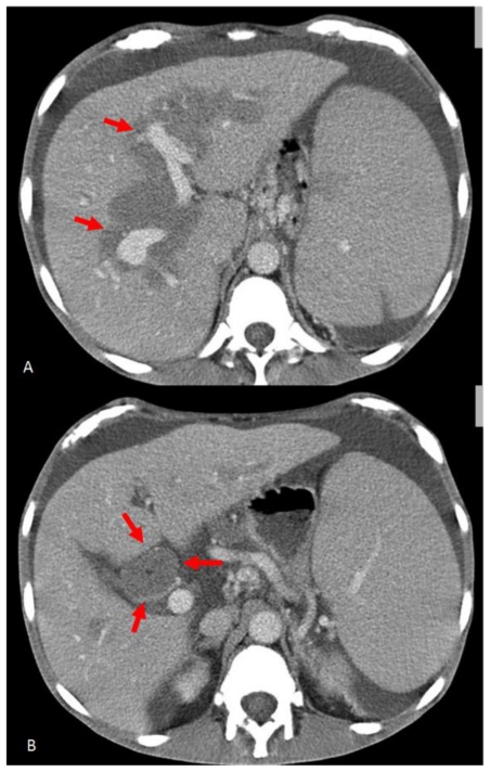
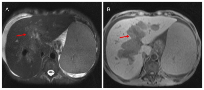
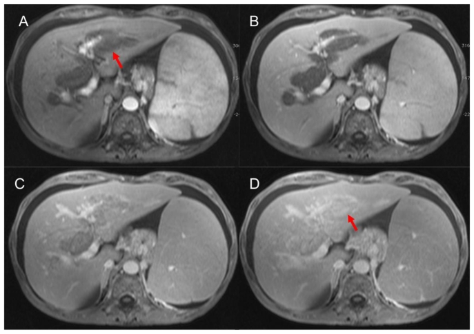
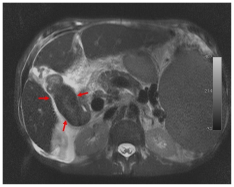
Extramedullary haematopoiesis is the production of blood elements outside the bone marrow cavity. In our case computed tomography and magnetic resonance imaging revealed the presence of a rare localization of extramedullary haematopoiesis with encasement of the biliary system in a 59 years-old male Caucasian patient, with chronic myelofibrosis and hepatic failure's symptomatology. Computed tomography detected the presence of homogeneous hypodense tissue around intra-hepatic bile ducts with minimal contrast enhancement, strongly suggestive for extramedullary haematopoiesis. Magnetic resonance confirmed the presence of a solid tissue surrounding the biliary tree, showing late enhancement after gadolinium administration suggestive for non-active lesion of extramedullary haematopoiesis. Final diagnosis was established by percutaneous biopsy.

Keywords: Extramedullary haematopoiesis; MRI; biliary system; computed tomography; diagnostic imaging; magnetic resonance imaging.


Figures

Figure 1
Figure 1
59 years-old male Caucasian patient with extramedullary haematopoiesis. Abdominal ultrasonography (GE Healthcare, convex probe at 4.0 MHz) evidenced near the hilum of the liver, above the portal vein, solid tissue, with irregular margin, hypoechoic to the surrounding liver (red arrow).
Figure 2
Figure 2
59 years-old male Caucasian patient with extramedullary haematopoiesis. Contrast-enhanced computed tomography scans (Sensation 16 Siemens, Erlangen, 332 mA, 120 KV, slice thickness 1–5 mm) (Portal phase: acquisition after 60 s non-ionic iodinated i.v. contrast injection) of the upper abdomen (non-ionic iodinated contrast medium: Visipaque GE Healthcare 320 mg I/ml, 100 ml, flow 2 ml/s). Axial CT images demonstrate homogeneous hypo-attenuating soft tissue around intra-hepatic bile ducts (red arrows) (A), with minimal enhancement, encasing the common bile duct (red arrows) (B).
Figure 3
Figure 3
59 years-old male Caucasian patient with extramedullary haematopoiesis. Magnetic resonance imaging of the upper abdomen (Symphony Siemens 1.5 T, Erlangen). A: Axial T2-weighted images TSE fat sat (TE: 84 ms, TR: 111060 ms; slice thickness: 5.0 mm) the pathological tissue shows inhomogeneous hyperintense signal (red arrow). B: Axial T1-weighted image, 3D-GRE, (TR= 5.12 ms, TE= 2.51 ms, slice thickness= 3.5 mm): the pathological tissue shows hypointense signal (red arrow).
Figure 4
Figure 4
59 years-old male Caucasian patient with extramedullary haematopoiesis. Magnetic resonance imaging of the upper abdomen (Symphony Siemens 1.5 T, Erlangen). Axial T1-weighted image, 3D-GRE, (TR= 5.12 ms, TE= 2.51 ms, slice thickness= 3.5 mm): plain acquisition in which extramedullary haematopoiesis appears hypointense to liver parenchyma; after intravenous administration of gadolinium-based contrast agent (Gd-BOPTA; Multihance, Bracco, Italy; 0,1 mmol/Kg; flow 2 ml/s) on scans acquired in arterial (A) and portal (B) phases the lesion reveal minimal enhancement. The pathological tissue shows progressive enhancement 5 (C) and 15 (D) minutes after i.v. injection (red arrows).
Figure 5
Figure 5
59 years-old male Caucasian patient with extramedullary haematopoiesis. Magnetic resonance imaging of the upper abdomen (Symphony Siemens 1.5 T, Erlangen). Axial T2-weighted images TSE fat sat (TE: 84 ms, TR: 111060 ms; slice thickness: 5.0 mm ): the usual hyperintense T2-w gallbladder signal was fully replaced by slightly high signal of pathological tissue (black arrows).

References

    1. Lichtman M, Beutler E, Kaushansky K, et al. Williams Hematology. 7th edn. New York: McGraw-Hill; 2005.
    1. Siniluoto TM, Hyvärinen SA, Päivänsalo MJ, Alavaikko MJ, Suramo IJ. Abdominal ultrasonography in myelofibrosis. Acta Radiol. 1992;33:343–346. - PubMed
    1. Warshauer DM, Schiebler ML. Intrahepatic extramedullary hematopoiesis: MR, CT and sonographic appearance. J Comput Assist Tomogr. 1991;15:683–685. - PubMed
    1. Dewar G, Leung NW, Ng HK, Bradley M, Li AK. Massive, solitary, intrahepatic, extramedullary hematopoietic tumor in thalassemia. Surgery. 1990 Jun;107(6):704–7. - PubMed
    1. Kumar A, Aggarwal S, de Tilly LN. Thalassemia major with extramedullary hematopoiesis in the liver. Semin Roentgenol. 1995;30(2):99–101. - PubMed

LinkOut - more resources