. 2010;4(3):31-4.
doi: 10.3941/jrcr.v4i3.352. Epub 2010 Mar 1.

FDG uptake in sternoclavicular joint synovitis: mimic of internal mammary adenopathy

Affiliations

FDG uptake in sternoclavicular joint synovitis: mimic of internal mammary adenopathy

Eugene Lin. J Radiol Case Rep. 2010.

Abstract

False-positive FDG uptake has been noted in a wide range of benign processes. In this report, we describe a case of FDG uptake in unilateral sternoclavicular synovitis which mimicked an internal mammary node in appearance. Knowledge of this potential false-positive is particularly important in breast cancer patients with a propensity of internal mammary nodal metastases.

Keywords: Positron emission tomography; arthritis; breast cancer; fluorodeoxyglucose; sternoclavicular joint; synovitis.


Figures

Figure 1
Figure 1
60 year old woman with a history of breast cancer and sternoclavicular joint synovitis mimicking internal mammary adenopathy. FDG PET/CT was performed 60 minutes after the injection of 19.8 mCi of FDG. Three axial images from this study are provided from superior (top) to inferior (bottom). This demonstrates soft tissue posterior to the right sternum with focal FDG uptake. However, the superior aspect of this soft tissue extends to the sternoclavicular joint (arrowhead). Another clue that this may not be internal mammary adenopathy is that the internal mammary nodes are typically just lateral to the sternum, while this tissue is slightly more medial. In addition, there is also soft tissue on the left side (arrow); however, the right-sided soft tissue is larger.
Figure 2
Figure 2
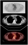
60 year old woman with a history of breast cancer and sternoclavicular joint synovitis mimicking internal mammary adenopathy. PET, CT, and fused PET/CT images from the same study as figure 1 (FDG PET/CT was performed 60 minutes after the injection of 19.8 mCi of FDG.), demonstrate the FDG uptake on the PET image (arrow) which fuses to soft tissue adjacent to the sternoclavicular joint on CT.
Figure 3
Figure 3
60 year old woman with a history of breast cancer and sternoclavicular joint synovitis mimicking internal mammary adenopathy. Bone scan performed 4 hours after the injection of 24.2 mCi of Tc-99m methylene diphosphonate demonstrates increased uptake in the right sternoclavicular joint (arrow).
Figure 4
Figure 4
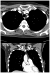
60 year old woman with a history of breast cancer and sternoclavicular joint synovitis mimicking internal mammary adenopathy. Axial and coronal reconstructed image from a CT (General Electric Lightspeed VCT, kVp 120, mAs 266, 5 mm reconstruction) performed with 125 cc of Isovue-370 contrast demonstrates peripheral enhancement of the right-sided soft tissue (arrows) noted on the prior FDG PET/CT. On this study, the peripheral enhancement demarcates the area of soft tissue, which is seen to arise from the sternoclavicular joint.

References

    1. Gorospe L, Raman S, Echeveste J, et al. Whole-body PET/CT: spectrum of physiological variants, artifacts and interpretative pitfalls in cancer patients. Nucl Med Commun. 2005;26(8):671–87. - PubMed
    1. Basu S, Tiwari BP. Asymmetric 18F-FDG uptake in the infradiaphragmatic brown adipose tissue (BAT) mimicking adrenal metastasis: A relatively rare site of brown fat and a potential source for false-positive FDG study. Radiology Case. 2009 Oct;3(10):19–22. - PMC - PubMed
    1. Veronesi U, Cascinelli N, Greco M, et al. Prognosis of breast cancer patients after mastectomy and dissection of internal mammary nodes. Ann Surg. 1985;202:702–7. - PMC - PubMed
    1. Bellon JR, Livingston RB, Eubank WB, et al. Evaluation of the internal mammary lymph nodes by FDG-PET in locally advanced breast cancer (LABC) Am J Clin Oncol. 2004;27(4):407–10. - PubMed
    1. Eubank WB, Mankoff DA, Takasugi J, et al. 18 fluorodeoxyglucose positron emission tomography to detect mediastinal or internal mammary metastases in breast cancer. J Clin Oncol. 2001;19(15):3516–23. - PubMed

LinkOut - more resources