. 2010;4(4):20-4.
doi: 10.3941/jrcr.v4i4.360. Epub 2010 Apr 1.

Stent-assisted coil embolization of a wide-necked renal artery aneurysm

Affiliations

Stent-assisted coil embolization of a wide-necked renal artery aneurysm

Bjoern Kitzing et al. J Radiol Case Rep. 2010.

Abstract

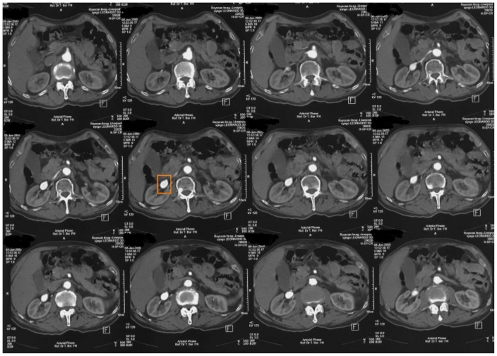
We present a case of stent-assisted coil embolization of a wide-necked renal artery aneurysm performed at our institution. The technique involved a stent being delivered over the neck of the aneurysm. Subsequently a catheter was placed into the aneurysm through the stent mesh and the aneurysm was then filled with detachable coils. Complete aneurysm occlusion was obtained and there was no evidence to suggest renal infarction on a follow-up contrast CT scan 6 months later. Our preliminary experience suggests that stent-assisted coil embolization of wide-necked renal artery aneurysms is a technically challenging but potentially effective renal-sparing endovascular approach.

Keywords: Renal artery aneurysm; coil embolization; stent.


Figures

Figure 1
Figure 1
Axial images of contrast computed tomography of a 77-year-old male with hypertension showing an oval right renal artery aneurysm measuring 2.5cm in diameter (highlighted by orange rectangle).
Figure 2
Figure 2
Axial image of contrast computed tomography of a 77-year-old male with hypertension showing an oval right renal artery aneurysm measuring 2.5cm in diameter (highlighted by orange rectangle).
Figure 3
Figure 3
Maximum intensity projection (MIP) images of a 77-year-old male with hypertension demonstrating an oval right renal artery aneurysm measuring 2.5cm in diameter (highlighted by orange rectangle).
Figure 4
Figure 4
Abdominal angiogram of a 77-year-old male with hypertension showing an oval aneurysm measuring approximately 2.5cm in maximum diameter arising from the upper first divisional vessel of the right renal artery.
Figure 5
Figure 5
Renal angiography of a 77-year-old male with hypertension demonstrating a catheter positioned through the stent in the upper first divisional vessel of the right renal artery and deployment of the first coil within the aneurysm.
Figure 6
Figure 6
Renal angiography of a 77-year-old male with hypertension showing multiple coils filling the right renal artery aneurysm after stent-assisted coil deployment.
Figure 7
Figure 7
Axial image of contrast computed tomography of a 77-year-old male 6 months after stent-assisted coil embolization of a right renal artery aneurysm showing no evidence to suggest renal infarction.

References

    1. Rouppe DL. Nova Acta Phys-Med Acad Nat Curios. 1770;iv:76.
    1. Stanley JC, Rhodes EL, Gewertz BL, et al. Renal artery aneurysms. Arch Surg. 1975;110:1327–1333. - PubMed
    1. Tham G, Ekelund L, Herrlin K, et al. Renal artery aneurysms. Natural history and prognosis. Ann Surg. 1983 Mar;197(3):348–52. - PMC - PubMed
    1. Dzsinich C, Gloviczki P, McKusick MA, et al. Surgical management of renal artery aneurysm. Cardiovasc Surg. 1993 Jun;1(3):243–7. - PubMed
    1. Lumsden AB, Salam TA, Walton KG. Renal artery aneurysm: a report of 28 cases. Cardiovasc Surg. 1996 Apr;4(2):185–9. - PubMed

LinkOut - more resources