

doi: 10.3941/jrcr.v4i6.439.
Epub 2010 Jun 1.
Giant aneurysm formation in sporadic renal angiomyolipoma
Affiliations
- PMID: 22470737
- PMCID: PMC3303414
- DOI: 10.3941/jrcr.v4i6.439
Item in Clipboard
Giant aneurysm formation in sporadic renal angiomyolipoma
J Radiol Case Rep.
2010.
Abstract
Angiomyolipomas are the most common mesenchymal renal neoplasms. Two types have been described: (i) sporadic angiomyolipoma and (ii) angiomyolipoma associated with tuberous sclerosis. Giant aneurysm formation is usually noted in angiomyolipomas associated with tuberous sclerosis and is rare in sporadic variety. Tumor diameter and aneurysm diameter have been used as predictors of rupture. We report a rare case of aneurysm formation in a sporadic angiomyolipoma.
Keywords: Aneurysm; Angiomyolipoma; Computed Tomography; Tuberous Sclerosis complex.
Figures
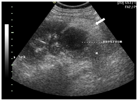
42 yr female with aneurysm formation in right renal angiomyolipoma: Transabdominal ultrasonography using 3.5 MHz electrical curvilinear array probe (Philips HDI 4000) of right lumbar region shows 8 × 7 × 8.5 cm heteroechoic mass in lower pole. The mass (m) shows central anechoic region (marked aneurysm) and a thick hyperechoic periphery (arrow)

42 yr female with aneurysm formation in right renal angiomyolipoma: Transabdominal ultrasonography using 3.5 MHz electrical curvilinear array probe (Philips HDI 4000) of right lumbar region with application of color Doppler shows “swirling” flow pattern within the anechoic region of the mass (arrow).
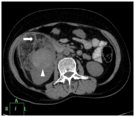
42 yr female with aneurysm formation in right renal angiomyolipoma: Plain axial CT scan (Philips 40 slice multidetector helical CT, at lumbar level) shows mixed density right renal mass with areas of fat attenuation (arrow). Few areas of hyperdensity were also noted within the mass (arrowhead). Parameters used: kVp - 120; mA - 306; slice thickness - 3mm.
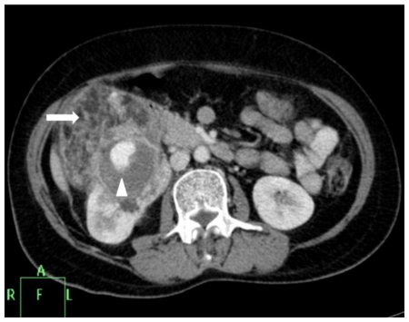
42 yr female with aneurysm formation in right renal angiomyolipoma: Contrast enhanced axial CT (Philips 40 slice multidetector helical CT at same level in arterial phase using non-ionic iodinated contrast agent Iohexol 90 ml manually injected) shows moderate heterogenous enhancement of the mass along with septal enhancement (arrow head). Intense vascular equivalent enhancing area is seen within mass consistent with partially thrombosed aneurysm (arrow). Parameters used: kVp - 120; mA - 206; slice thickness - 2mm.
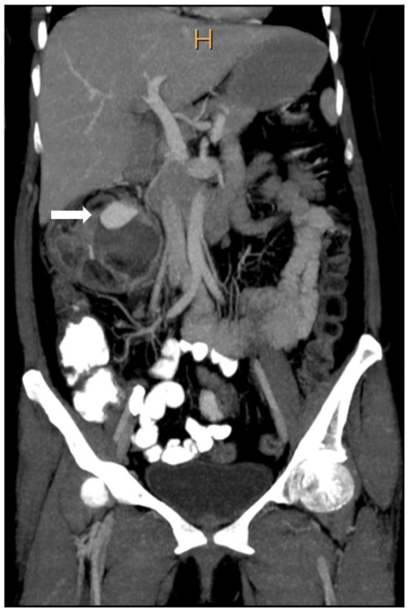
42 yr female with aneurysm formation in right renal angiomyolipoma: Coronal reformatted CT in maximum intensity projection (Philips 40 slice multidetector helical CT in arterial phase using non-ionic iodinated contrast agent Iohexol 90 ml manually injected) clearly demonstrates fatty right lower pole renal mass with partially thrombosed aneurysm (arrow). Parameters used: Parameters used: kVp - 120; mA - 206; Maximum Intensity Projection applied.
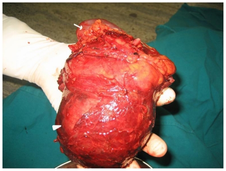
42 yr female with right renal angiomyolipoma: Post operative appearance of the radical nephrectomy specimen showing lower pole mass (arrowhead) and the normal renal parenchyma (arrow).
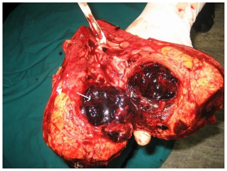
42 yr female with right renal angiomyolipoma: Cut section of the radical nephrectomy specimen showing fat containing 9.5 × 9 × 8 cm mass (arrowhead). Arrow points to aneurysm with thrombosed blood within.
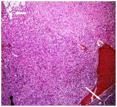
42 yr female with right renal angiomyolipoma: Histopathology with H & E staining (low magnification) showing mature adipose cells (arrow) and areas of hemorrhage (curved arrow). Long straight arrow shows the normal renal tissue.
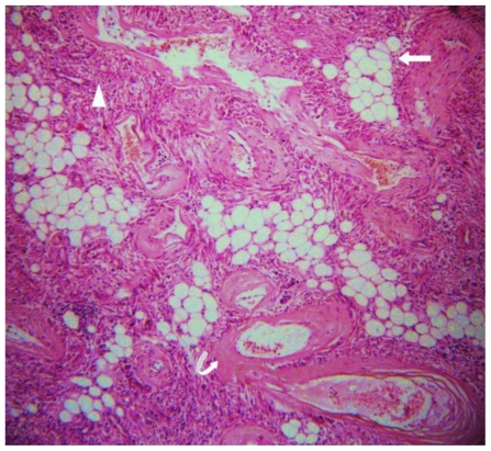
42 yr female with right renal angiomyolipoma: Histopathology with H & E staining (high magnification) showing admixture of mature adipose tissue (arrow), thick walled blood vessels lacking elastic tissue (curved arrow) and smooth muscle cells (arrowhead).
References
-
- Fujii Y, Ajima J, Oka K, et al. Benign renal tumors detected among healthy adults by abdominal ultrasonography. Eur Urol. 1995;27:124–127. - PubMed
-
- Casper Keith A, Donnelly Lane F, Chen Bin, Bissler John J. Tuberous Sclerosis Complex: Renal imaging findings. Radiology. 2002;225:451–456. - PubMed
-
- Evans JC, Curtis J. The radiological appearances of tuberous sclerosis. Br J Radiol. 2000;73:865, 91–98. - PubMed
-
- Pea M, Bonetti F, Martignoni G, et al. Apparent renal cell carcinomas in tuberous sclerosis are heterogeneous: the identification of malignant epithelioid angiomyolipoma. Am J Surg Pathol. 1998;22:80–187. - PubMed
LinkOut - more resources
Full Text Sources