. 2010;4(8):22-8.
doi: 10.3941/jrcr.v4i8.487. Epub 2010 Aug 1.

Extramedullary duodenal plasmacytoma presenting with gastric outlet obstruction and painless jaundice

Affiliations

Extramedullary duodenal plasmacytoma presenting with gastric outlet obstruction and painless jaundice

Adib R Karam et al. J Radiol Case Rep. 2010.

Abstract

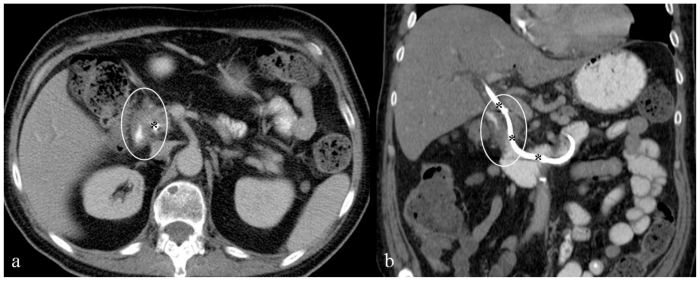
Malignant plasma cells in multiple myeloma are predominantly confined to the medullary space of the skeletal system, therefore the disease course will be dominated by signs and symptoms related to bone marrow infiltration and destructive bone lesions with their consequences as well as abnormal protein production. Visceral extramedullary plasmacytoma involving the gastrointestinal system and particularly the duodenum is a rare manifestation of the disease. We report a case of duodenal extramedullary plasmacytoma presenting with gastric outlet obstruction and painless jaundice, in a patient treated for multiple myeloma. Diagnosis was first suggested on imaging, and proved by endoscopic biopsy. The duodenal mass resolved following chemotherapy.

Keywords: Multiple myeloma; duodenal plasmacytoma; extramedullary multiple myeloma; gastric outlet obstruction; gastrointestinal involvement; painless jaundice.


Figures

Figure 1
Figure 1
66-year-old male patient presented with severe nausea and vomiting in the setting of a two-week history of worsening fatigue, pruritus, and jaundice. Axial (a) and coronal (b) contrast enhanced (IV and PO) CT images through the mid abdomen obtained in the equilibrium phase show a large solid enhancing soft tissue tumor, measuring approximately 4.4 × 6.0 × 5.8 cm. It appears originating from the medial wall of the descending portion of the duodenum narrowing its lumen (asterisk), involving the medial aspect of the common bile duct and pancreatic head, resulting in significant dilatation of the intrahepatic (thin white arrows) and extrahepatic (thick white arrows) biliary tree; the pancreatic duct is also dilated (not shown). Enlarged regional mesenteric lymph nodes were noted (black cursor). Note significant fluid residue in the stomach. GB: gallbladder; T: tumor; P: pancreatic head; S: stomach. [Technique: KVp = 120; mA = 356; Slice Thickness = 4.00 mm; Dose of intravenous contrast: Iopamidol (Isovue-300), 100 ml].
Figure 2
Figure 2
66-year-old male patient presented with severe nausea and vomiting in the setting of a two-week history of worsening fatigue, pruritus, and jaundice. Single three-dimensional Radial MRCP Breath Hold image (TR/TE = 2857.85 msec./1027.58 msec. flip angle = 90.0 degrees) demonstrates significant intra- and extrahepatic biliary ductal dilatation as well as main pancreatic duct dilatation. There is a large, non-cystic, space occupying mass (orthogonal white arrows) impinging on the duodenal lumen. Noted is significant fluid residue in the stomach. B: biliary tree; PD: pancreatic duct; D: duodenum; S: stomach.
Figure 3
Figure 3
66-year-old male patient presented with severe nausea and vomiting in the setting of a two-week history of worsening fatigue, pruritus, and jaundice. a: Transverse noncontrast, T2-weighted, fat-suppressed image (TR/TE = 2600 msec./160 msec.) demonstrates the duodenal tumor showing homogenous high signal intensity. b: Transverse noncontrast, T1-weighted, 2D fat- suppressed spoiled gradient-echo sequence (TR/TE = 177 msec./4.3 msec.; flip angle = 80 degrees) demonstrates the duodenal tumor showing homogenous signal intensity isointense to the paraspinal muscles. c–f: Transverse, T1-weighted, fast-suppressed, 3D spoiled gradient echo (LAVA) sequences (TR/TE = 4.216 msec./2.02 msec. flip angle = 12 degrees), before and after intravenous contrast (20 ml gadobenate dimeglumine, Multihance). Duodenal tumor demonstrates mild enhancement during the hepatic arterial-dominant phase (d), which becomes homogenous and more intense after one minute (e), and two-minute delay (f). T: duodeal tumor.
Figure 4
Figure 4
66-year-old male patient presented with severe nausea and vomiting in the setting of a two-week history of worsening fatigue, pruritus, and jaundice. a, b: endoscopic images from the first and second segments of the duodenum showing an extrinsic compression on the medial wall of the duodenum (T) slightly narrowing the duodenal lumen (curved white arrows). c: Endoscopic image from the periampullary region showing mucosal ulceration (asterisks) and bleeding (B), overlying the extrinsic impression from the tumor (T).
Figure 5
Figure 5
66-year-old male patient presented with severe nausea and vomiting in the setting of a two-week history of worsening fatigue, pruritus, and jaundice. Axial (a) and coronal (b) contrast enhanced (IV and PO) CT images through the mid abdomen obtained in the equilibrium phase, six weeks following chemotherapy, show resolution of the duodenal soft tissue tumor; there remains minimal circumferential duodenal wall thickening (white cursors). Noted is a biliary stent (asterisks) seen from the level of the porta hepatis across the ampulla of Vater, its distal tip formed in the fourth segment of the duodenum. [Technique: KVp = 120; mA = 275; Slice Thickness = 5.00 mm; Dose of intravenous contrast: Iopamidol (Isovue-300), 100 ml].

References

    1. Pimmental R, vanstolk R. Gastric plasmacytoma: A rare cause of massive gastrointestinal bleeding. Am J Gastroenterol. 1993;88:1963–1964. - PubMed
    1. Chim CS, Wong WM, Nicholls J, Chung LP, Liang R. Extramedullary sites of invovelment in hematologic malignancies: case3. Hemorrhagic gastric plasmacytoma as the primary presentation in multiple myeloma. J Clin Oncol. 2002;20:344–347. - PubMed
    1. Alexiou C, Kau RJ, Dietzfelbinger H, et al. Extramedullary plasmacytoma: tumor occurrence and therapeutic concepts. Cancer. 1999;85:2305–2314. - PubMed
    1. Dolin S, Dewer J. Extramedullary plasmacytoma. Am J Pathol. 1955;32:83–103. - PMC - PubMed
    1. Goldstein WB, Poker N. Multiple myeloma involving the gastrointestinal tract. Gastroenterology. 1966;51:87–93. - PubMed

LinkOut - more resources