Case Reports
. 2011;5(6):18-24.
doi: 10.3941/jrcr.v5i6.682. Epub 2011 Jun 1.

Plunging ranula

Affiliations
Case Reports

Plunging ranula

Vivek Kalra et al. J Radiol Case Rep. 2011.

Abstract

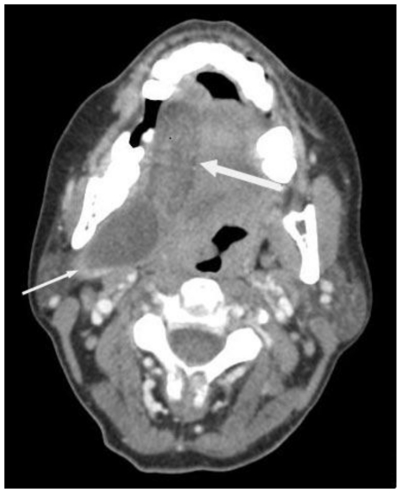
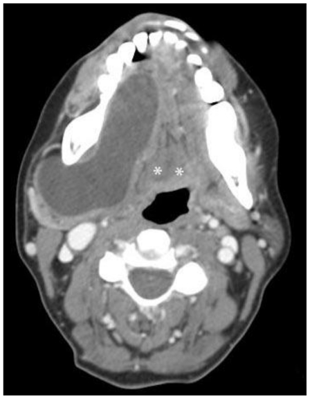
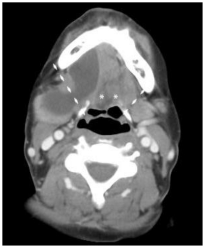
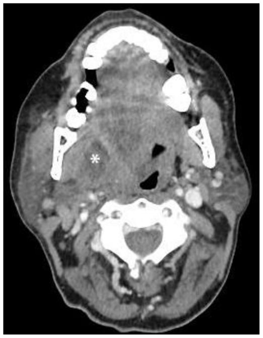
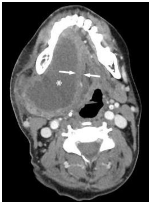
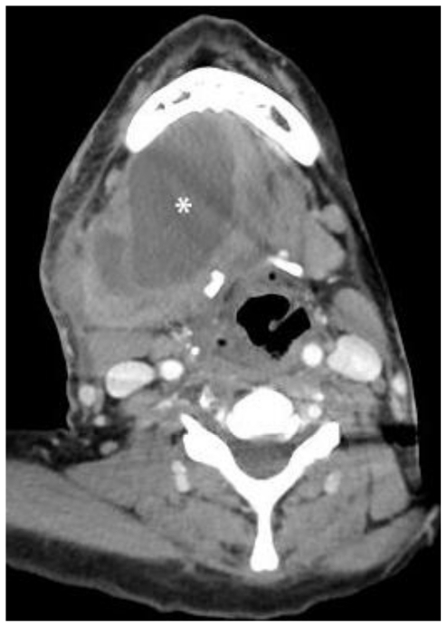
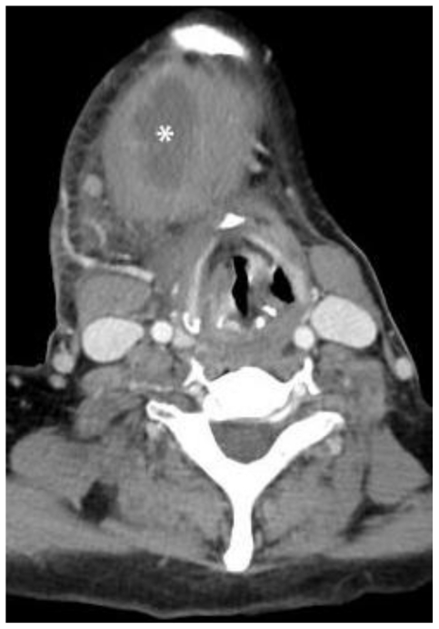
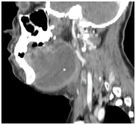
Plunging ranulas are rare cystic masses in the neck that are mucous retention pseudocysts from an obstructed sublingual gland. They "plunge" by extending inferiorly beyond the free edge of the mylohyoid muscle, or through a dehiscence of the muscle itself, to enter the submandibular space. Imaging demonstrates a simple cystic lesion in the characteristic location and can be used to delineate relevant surgical anatomy. Surgical excision of the collection and the involved sublingual gland is performed for definitive treatment. We present a case of plunging ranula in a 44 year old female who presented with a painless, slowly enlarged neck mass. Plunging ranulas should be considered in the differential diagnosis of cystic neck masses, specifically when seen extending over, or through, the mylohyoid muscle.

Keywords: cystic neck masses; diving ranula; plunging ranula; ranula.


Figures

Figure 1
Figure 1
Initial contrast enhanced axial Computed Tomography - 100 cc of Omnipaque-300 (KVp 120, mA 20 * 3572ms). 44-year-old female with plunging ranula - Superior aspect of the lesion - Shows a non-enhancing, water-density mass from the right sublingual space displacing the tongue to the left. A large component of the mass extends posterolaterally into the submandibular space (small arrow). Mass effect can be appreciated with displacement of the parapharyngeal and mucosal pharyngeal spaces. The lesion measures 6.7 × 2.2 × 4.5 cm and is of water-density. It is non-enhancing, homogenous, smoothly-marginated, and without internal septations. Smooth tapering anteriorly into the sublingual space, forming the “tail sign,” is seen, but better appreciated on stack images (large arrow).
Figure 2
Figure 2
Initial contrast enhanced axial Computed Tomography - 100 cc of Omnipaque-300 (KVp 120, mA 20 * 3572ms). 44-year-old female with plunging ranula. Slightly inferior slice through the mid-portion of the lesion - shows that the anterior aspect of the lesion is centered in the sublingual space, lateral to the geniohyoid muscle (left *). The contralateral geniohyoid (right *) is also seen. Extension into the sublingual, parapharyngeal, and submandibular spaces is seen. The lesion measures 6.7 × 2.2 × 4.5 cm and is of water-density density. It is non-enhancing, homogenous, smoothly-marginated, and without internal septations.
Figure 3
Figure 3
Initial contrast enhanced axial Computed Tomography - 100 cc of Omnipaque-300 (KVp 120, mA 20 * 3572ms). 44-year-old female with plunging ranula. A more inferior level, at the level of the hyoid bone. The geniohyoids (labeled with “*”) can be appreciated inserting on the hyoid bone. The dashed lines show the posterolateral free-margins of the mylohyoid muscle. The lesion measures 6.7 × 2.2 × 4.5 cm and is of water-density. It is non-enhancing, homogenous, smoothly-marginated, and without internal septations. The posterior aspect of the lesion can be seen extending posteriorly over the approximate location of the free edge of the mylohyoid bone.
Figure 4
Figure 4
Follow-up contrast enhanced axial Computed Tomography performed 2 months later - 80 cc of Omnipaque-300 (KVp 120, mA 20 * 3575 ms) 44-year-old female with infected plunging ranula. Superior aspect of the lesion (labeled with *). The mass has unchanged anatomic relationships, but has slightly enlarged and now measures 6.9 × 3.7 × 4.0 cm. It remains homogenous, smoothly-marginated, and without internal septations. Thick rim-enhancement is now seen, consistent with secondary infection. Mass effect with tracheal deviation and compression is worse and there is new right submandibular and cervical reactive lymphadenopathy. The mass was surgically excised and pathologically proven to be a plunging ranula.
Figure 5
Figure 5
Follow-up contrast enhanced axial Computed Tomography performed 2 months later - 80 cc of Omnipaque-300 (KVp 120, mA 20 * 3575 ms). 44-year-old female with infected plunging ranula. Slightly more caudal, the mid-portion of the lesion (labeled with *). The mass has unchanged anatomic relationships, but has slightly enlarged and now measures 6.9 × 3.7 × 4.0 cm. Smooth tapering anteriorly into the sublingual space, forming the “tail sign,” is again seen, but better appreciated on stack images. It remains homogenous, smoothly-marginated, and without internal septations. Thick rim-enhancement is now seen (labeled with arrows), consistent with secondary infection. Mass effect with tracheal deviation and compression is worse and there is new right submandibular and cervical reactive lymphadenopathy. The mass was surgically excised and pathologically proven to be a plunging ranula.
Figure 6
Figure 6
Follow-up contrast enhanced axial Computed Tomography performed 2 months later - 80 cc of Omnipaque-300 (KVp 120, mA 20 * 3575 ms). 44-year-old female with infected plunging ranula. More caudal, at the level of hyoid bone. The mass (labeled with *) has unchanged anatomic relationships, but has slightly enlarged and now measures 6.9 × 3.7 × 4.0 cm. It remains homogenous, smoothly-marginated, and without internal septations. Thick rim-enhancement is now seen, consistent with secondary infection. Mass effect with tracheal deviation and compression is worse and there is new right submandibular and cervical reactive lymphadenopathy. The mass was surgically excised and pathologically proven to be a plunging ranula.
Figure 7
Figure 7
Follow-up contrast enhanced axial Computed Tomography performed 2 months later - 80 cc of Omnipaque-300 (KVp 120, mA 20 * 3575 ms). 44-year-old female with infected plunging ranula. The image demonstrates the inferior aspect of the lesion. The mass (labeled with *) has unchanged anatomic relationships, but has slightly enlarged and now measures 6.9 × 3.7 × 4.0 cm. It remains homogenous, smoothly-marginated, and without internal septations. Thick rim-enhancement is now seen, consistent with secondary infection. Mass effect with tracheal deviation and compression is worse and there is new right submandibular and cervical reactive lymphadenopathy. The mass was surgically excised and pathologically proven to be a plunging ranula.
Figure 8
Figure 8
Follow-up contrast enhanced Computed Tomography performed 2 months later - 80 cc of Omnipaque-300 (KVp 120, mA 20 * 3575 ms). 44-year-old female with plunging ranula. Sagittal reconstruction to the right of midline. The mass (labeled with *) has unchanged anatomic relationships, but has slightly enlarged and now measures 6.9 × 3.7 × 4.0 cm. It remains homogenous, smoothly-marginated, and without internal septations. Thick rim-enhancement is now seen, consistent with secondary infection. Mass effect with tracheal deviation and compression is worse and there is new right submandibular and cervical reactive lymphadenopathy. The mass was surgically excised and pathologically proven to be a plunging ranula.

References

    1. Coit WE, Harnsberger HR, Osborn AG, Smoker WR, Stevens MH, Lufkin RB. Ranulas and their mimics: CT evaluation. Radiology. 1987 Apr;163(1):211–6. - PubMed
    1. Davison MJ, Morton RP, McIvor NP. Plunging Ranula: clinical observations. Head Neck. 1998 Jan;20(1):63–8. - PubMed
    1. Morton RP, Ahmad Z, Jain P. Plunging ranula: congenital or acquired. Otolaryngol Head Neck Surg. 2010 Jan;142(1):104–7. - PubMed
    1. Charnoff SK, Carter BL. Plunging Ranula: CT diagnosis. Radiology. 1986 Feb;158(2):467–8. - PubMed
    1. Macdonald AJ, Salzman KL, Harnsberger HR. Giant ranula of the neck: differentiation from cystic hygroma. AJNR Am J Neuroradiol. 2003 Apr;24(4):757–61. - PMC - PubMed

Publication types

LinkOut - more resources