

Vasa previa
J Prenat Med.
2007 Jan.
No abstract available
Figures

Drawing showing the inner view to the uterus, towards the cervix, demonstrating the anatomical relations in case of succenturiate placenta. The vessels between the main and succenturiate lobe are crossing the inner cervical os.

Drawing showing the inner view to the uterus, towards the cervix, demonstrating the anatomical relations in case of velamentous insertion of the umbilical cord.
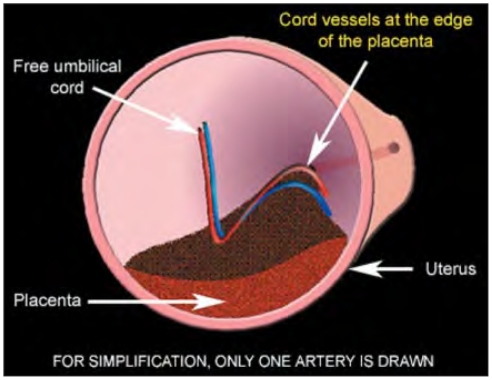
Drawing showing the inner view to the uterus, towards the cervix, demonstrating the anatomical relations in case of marginal placenta with vessels running at the edge of placenta and crossing the inner cervical os. By trophotropism, the marginal edge of the placenta regresses, leaving the vessel in front of the inner cervical os.

Pathological specimen shows the fetal side of bilobate placenta with velamentous insertion of the umbilical cord between the placental lobes. (Courtesy Francois Manson and TheFetus.net).

Pathological specimen shows the maternal side of the bilobate placenta. (Courtesy Francois Manson and TheFetus. net).

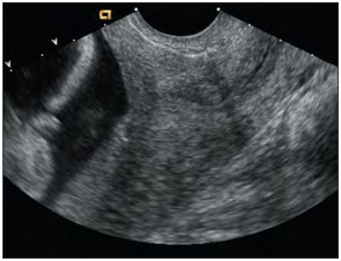
Second trimester vaginal 2D sonography shows a sagittal section through the cervix. In this gray scale mode no vessels are visible crossing the inner cervical os.

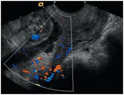
The same scan as in image 5 using color Doppler shows a vasa previa crossing the inner cervical os.
Second trimester vaginal 2D sonography shows a sagittal section through the cervix with the marginal placenta previa localized at the dorsal wall of the uterus.

The same scan as in image 3 using color Doppler showing a vessel crossing the inner cervical os (vasa previa).

Proposed diagnostic algorithm for the second-trimester detection of the vasa previa.
A second trimester vaginal 2D ultrasonographic scan shows sagittal section through the cervix with amniotic fluid above.

The same second trimester vaginal sonography as in figure 2 using color Doppler showing a flushing artefact, caused by the movement of the amniotic fluid during fetal movement, imitating vasa previa.

A second trimester vaginal 2D ultrasonographic scan shows sagittal section through the cervix with suspicious vessels crossing inner cervical os (arrow).

The same scan as in figure 15 using the color Doppler clearly show that the suspicious structure is without Doppler signal and thus is not a vessel.

Second trimester vaginal Doppler image shows a high frequency fetal hart rate at the level of vasa previa. This helps to distinguish vasa previa from maternal cervical vessels.
References
-
- Lobstein J. Archives de L’art des Accouchements 1801. Strasbourg: p. 320.
-
- Gianopoulos J, Carver T, Tomich PG, Karlman R, Gadwood K. Diagnosis of vasa previa with ultrasonography. Obstet Gynecol. 1987 Mar;69(3 Pt 2):488–91. - PubMed
-
- Oyelese KO, Turner M, Lees C, Campbell S. Vasa previa: an avoidable obstetric tragedy. Obstet Gynecol Surv. 1999 Feb;54(2):138–45. - PubMed
-
- Strassmann P. Placenta previa. Arch Gynecol. 1902;67:112.
-
- Oyelese Y, Chavez MR, Yeo L, Giannina G, Kontopoulos EV, Smulian JC, Scorza WE. Three-dimensional sonographic diagnosis of vasa previa. Ultrasound Obstet Gynecol. 2004 Aug;24(2):211–5. - PubMed
LinkOut - more resources
Full Text Sources