Review
. 2012 Feb;4(2):280-308.
doi: 10.3390/v4020280. Epub 2012 Feb 13.

Henipavirus mediated membrane fusion, virus entry and targeted therapeutics

Affiliations
Review

Henipavirus mediated membrane fusion, virus entry and targeted therapeutics

Deborah L Steffen et al. Viruses. 2012 Feb.

Abstract

The Paramyxoviridae genus Henipavirus is presently represented by the type species Hendra and Nipah viruses which are both recently emerged zoonotic viral pathogens responsible for repeated outbreaks associated with high morbidity and mortality in Australia, Southeast Asia, India and Bangladesh. These enveloped viruses bind and enter host target cells through the coordinated activities of their attachment (G) and class I fusion (F) envelope glycoproteins. The henipavirus G glycoprotein interacts with host cellular B class ephrins, triggering conformational alterations in G that lead to the activation of the F glycoprotein, which facilitates the membrane fusion process. Using the recently published structures of HeV-G and NiV-G and other paramyxovirus glycoproteins, we review the features of the henipavirus envelope glycoproteins that appear essential for mediating the viral fusion process, including receptor binding, G-F interaction, F activation, with an emphasis on G and the mutations that disrupt viral infectivity. Finally, recent candidate therapeutics for henipavirus-mediated disease are summarized in light of their ability to inhibit HeV and NiV entry by targeting their G and F glycoproteins.

Keywords: antibody; attachment glycoprotein; entry; ephrin-B2; ephrin-B3; fusion glycoprotein; immunotherapy; inhibitor; membrane fusion; paramyxovirus; receptor.


Figures
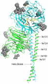
Figure 1
Figure 1
Model of the Hendra virus attachment G glycoprotein. The HeV-G ectodomain is shown in its dimer conformation. The secondary structure elements of the two globular head domains, colored in green and blue, are derived from the crystal structure, which also revealed the five predicted N-linked glycosylation sites (N306, N378, N417, N481 and N529) occupied by carbohydrate moieties (gray spheres) [46,47]. However, N378 was not modeled in the figure due to weak electron density. The G glycoprotein head domain folds as a six-blade β-propeller with disulfide bonds illustrated as yellow sticks. The residues of the ephrin-B2 G-H loop are shown in yellow. While the entire structure of the HeV-G stalk domain (residues 71–173) has not been determined, residues 77–136 are modeled for each monomer suggesting this region forms a discontinuous helix (Helix Break) [50]. The position of the HeV-G head dimer and stalks are oriented based on the alignment with the NDV structure and the receptor binding face of the blue monomer is facing out and the green monomer is facing left. Despite having two helical ranges, Thr-77 to Lys-95 and Thr-98 to Ser-135, the HeV-G stalk residues 98–135 appear equivalent to the HN glycoprotein stalk helix domain of the recently reported NDV structure [49]. Additionally, the Ile residues in the HeV-G stalk domain that can modulate conformational changes associated with receptor binding are indicated and are located in the alpha helical region of the HeV-G stalk domain that aligns with the NDV-HN stalk [51].
Figure 2
Figure 2
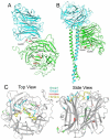
Mutations in HeV-G that affect fusion promoting activity. (A) Residues A257, A260, A443 and A465 that decrease HeV fusion when mutated are highlighted in red in the HeV-G globular head dimer. Based on the location of residues A257 and A260, it is likely they are necessary for interaction between the two globular heads, while residues A443 and A465 are centrally located in the globular head in a region of extensive hydrogen bonding. (B) The predicted structure of an HeV-G dimer with globular heads and stalk domains is shown with residues G449 and D468 highlighted in red. These two residues are located in proximity to the stalk domain, and mutation of these residues decreases HeV fusion, suggesting they may be involved in interactions between the globular heads and stalk domains that are essential for the fusion process. (C) Monomeric units of the globular head of HeV-G (gray) are shown in complex with the G-H loop residues FSPNLW of ephrin-B2 (yellow). Mutant Group 1 residues that decrease fusion are located further from the ephrin-B2 binding site and are shown in green. Mutant Group 2 residues that decrease fusion are shown in blue and cluster around the ephrinB2 binding site. Residues that enhance fusion (Group e1) are shown in red and are also distally located from the ephrin-B2 binding site. Given the divergent locations of the mutations that affect fusion, these mutations likely use different mechanisms, such as disrupting HeV-G structure or preventing ephrin-B2/B3 binding, to prevent HeV fusion.
Figure 3
Figure 3
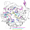
HeV-G residues that bind ephrin and monoclonal antibody m102.4. A monomeric unit of the globular head of HeV-G (gray) is shown with residues important for ephrin-B2 and -B3 binding highlighted in purple, residues required for binding mAb m102.4 shown in blue and the residues of the ephrin-B2 G-H loop in yellow. While the conformations of the residues may be slightly different, almost every residue involved in binding ephrin-B2 and -B3 is also required for binding m102.4, suggesting m102.4 prevents HeV and NiV infection by preventing ephrin-B2 and -B3 binding.

References

    1. Selvey L.A., Wells R.M., McCormack J.G., Ansford A.J., Murray K., Rogers R.J., Lavercombe P.S., Selleck P., Sheridan J.W. Infection of humans and horses by a newly described morbillivirus [see comments]. Med. J. Aust. 1995;162:642–645. - PubMed
    1. Chua K.B., Goh K.J., Wong K.T., Kamarulzaman A., Tan P.S., Ksiazek T.G., Zaki S.R., Paul G., Lam S.K., Tan C.T. Fatal encephalitis due to Nipah virus among pig-farmers in Malaysia [see comments]. Lancet. 1999;354:1257–1259. - PubMed
    1. Chua K.B., Bellini W.J., Rota P.A., Harcourt B.H., Tamin A., Lam S.K., Ksiazek T.G., Rollin P.E., Zaki S.R., Shieh W., et al. Nipah virus: A recently emergent deadly paramyxovirus. Science. 2000;288:1432–1435. - PubMed
    1. Eaton B.T., Broder C.C., Middleton D., Wang L.F. Hendra and Nipah viruses: Different and dangerous. Nat. Rev. Microbiol. 2006;4:23–35. - PMC - PubMed
    1. Chong H.T., Kunjapan S.R., Thayaparan T., Tong J., Petharunam V., Jusoh M.R., Tan C.T. Nipah encephalitis outbreak in Malaysia, clinical features in patients from Seremban. Can. J. Neurol. Sci. 2002;29:83–87. - PubMed

Publication types

MeSH terms

LinkOut - more resources