. 2012 May;168(2):165-77.
doi: 10.1111/j.1365-2249.2012.04567.x.

Tumour necrosis factor-mediated macrophage activation in the target organ is critical for clinical manifestation of uveitis

Affiliations

Tumour necrosis factor-mediated macrophage activation in the target organ is critical for clinical manifestation of uveitis

T K Khera et al. Clin Exp Immunol. 2012 May.

Abstract

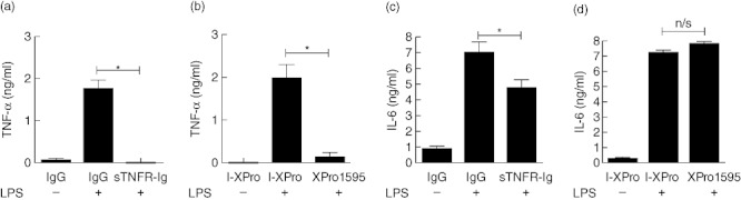
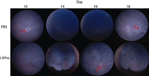
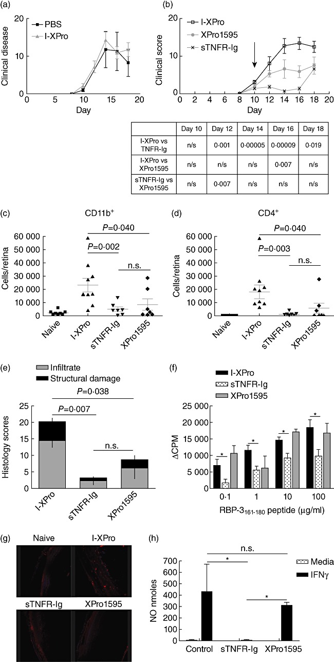
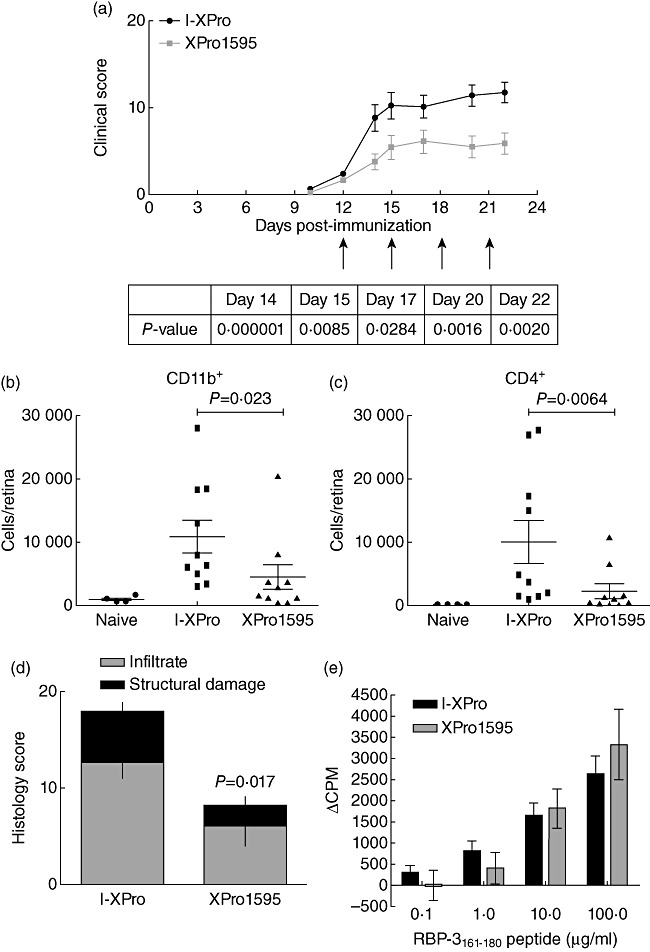
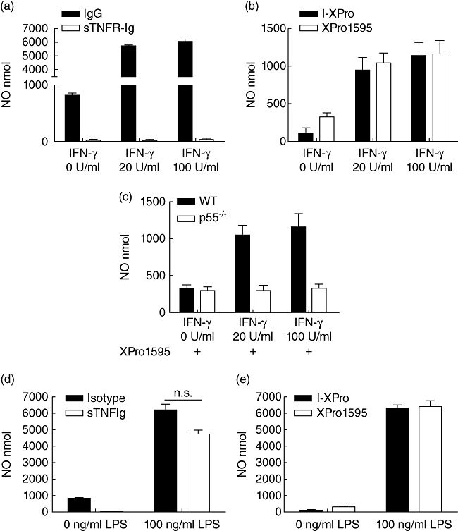
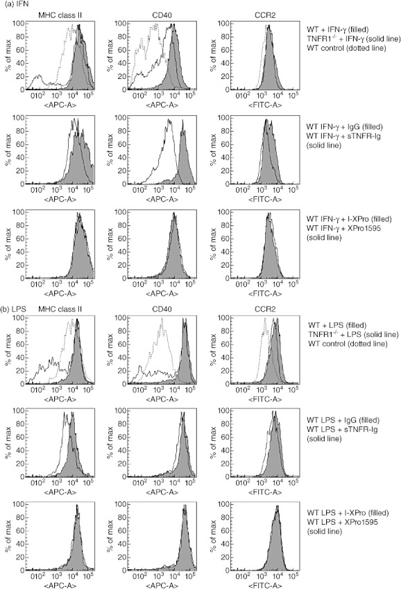
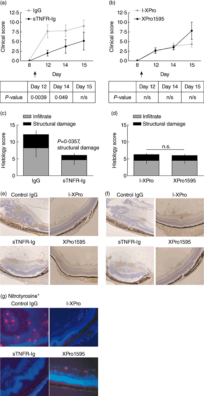
Clinically available anti-tumour necrosis factor (TNF) biologics, which inhibit both soluble (sTNF) and transmembrane forms (tmTNF) of TNF, eliminating all TNF signalling, have successfully treated autoimmune diseases including uveitis. These have potentially serious side effects such as reactivation of latent Mycobacterium tuberculosis and, therefore, more specific inhibition of TNF signalling pathways may maintain clinical efficacy while reducing adverse effects. To determine the effects of specific pharmacological inhibition of sTNF on macrophage activation and migration, we used a mouse model of uveitis (experimental autoimmune uveoretinitis; EAU). We show that selective inhibition of sTNF is sufficient to suppress EAU by limiting inflammatory CD11b(+) macrophages and CD4(+) T cell migration into the eye. However, inhibition of both sTNF and tmTNF is required to inhibit interferon-γ-induced chemokine receptor 2, CD40, major histocompatibility complex class II and nitric oxide (NO) up-regulation, and signalling via tmTNF is sufficient to mediate tissue damage. In confirmation, intravitreal inhibition of sTNF alone did not suppress disease, and inflammatory cells that migrated into the eye were activated, generating NO, thus causing structural damage to the retina. In contrast, intravitreal inhibition of both sTNF and tmTNF suppressed macrophage activation and therefore disease. We conclude that sTNF is required for inflammatory cell infiltration into target tissue, but at the tissue site inhibition of both sTNF and tmTNF is required to inhibit macrophage activation and to protect from tissue damage.


Figures

Fig. 1
Fig. 1
Both soluble tumour necrosis factor receptor-immunoglobulin (sTNFR-Ig) and XPro1595 can inhibit sTNF production but only sTNFR-Ig can suppress interleukin (IL)-6 secretion. Bone marrow-derived macrophages (BMDMϕ) from wild-type mice were incubated with 100 ng/ml lipopolysaccharide (LPS) for 24 h in the presence of 10 µg/ml human IgG or sTNFR-Ig (a,c) or 10 µg/ml I-XPro or XPro1595 (b,d). TNF (a,b) or IL-6 (c,d) production was quantified by enzyme-linked immunosorbent assay. Mean ± standard error shown, n = 3, *P < 0·05.
Fig. 2
Fig. 2
Topical endoscopic fundal imaging (TEFI). Experimental autoimmune uveoretinitis (EAU) was induced in B10.RIII mice, and on day 10 post-immunization (p.i.) the mice were treated intraperitoneally (i.p.) with 10 mg/kg [in 100 µl phosphate-buffered saline (PBS)] I-XPro or 100 µ1 PBS. Clinical progression of disease was assessed by TEFI from days 8 to 18; n = 4 mice per group and representative images are shown. In the PBS day 10 image, a raised and swollen optic disc is clearly visible (arrow 1). In the PBS-treated group on days 14 and 16, severe vitritis is causing a vitreous haze, precluding fundal imaging. On day 18 in the PBS-treated group, lesions (arrow 2) that correspond to histological retinal folds are present. In the I-XPro-treated mice on days 14 and 18, development of synechia was noted. On day 16 in the I-XPro-treated group, the optic disc is inflamed (raised and blurred margins; arrow 3) alongside widely distributed perivascular inflammation (vasculitis) and exudative retinal detachment as well as development of inflammatory retinal infiltrates.
Fig. 3
Fig. 3
Inhibition of soluble tumour necrosis factor (sTNF) can suppress experimental autoimmune uveoretinitis (EAU). EAU was induced in B10.RIII mice, and on day 10 post-immunization (p.i.) the mice were treated intraperitoneally (i.p.) with 10 mg/kg [in 100 µl phosphate-buffered saline (PBS)] I-XPro or 100 µ1 PBS. Clinical progression of disease with assessed by topical endoscopic fundus imaging (TEFI) from days 8 to 18; n = 4 mice per group (a). EAU was induced in B10.RIII mice, and on day 10 p.i. the mice were treated i.p. with 10 mg/kg of either I-XPro, sTNF receptor-immunoglobulin (sTNFR-Ig) or XPro1595 and the clinical progression of disease with assessed by TEFI from days 8 to 18. Treatment day is indicated by the arrow (b). The P-values for each day are tabulated below the graph. On day 18, the mice were killed and eyes taken for flow cytometric analysis of CD11b+ (c) and CD4+ (d) cells and histological analysis (e); mean ± standard error shown, n = 7–9 mice per group. On day 18 post-immunization, splenocytes were also used for a proliferation assay using 0·1–100 µg retinol-binding protein 3 (RBP-3)161–181 peptide (f); mean ± standard error shown. Eye sections from each mouse were also processed to detect nitrotryosine (red) and nuclear staining with 4′,6-diamidino-2-phenylindole (DAPI) (blue), ×20 magnification (g). Naive mice were injected (i.p.) with 200 µg/mouse of I-XPro, sTNFR-Ig or XPro1595 and after 3 days the spleens removed and macrophages isolated using magnetic affinity cell sorting (MACS). The macrophages were stimulated in vitro with 100 U/ml interferon (IFN)-γ for 72 h prior to nitric oxide (NO) quantification; n = 3 (h).
Fig. 4
Fig. 4
Inhibition of soluble tumour necrosis factor (sTNF) can also suppress established experimental autoimmune uveoretinitis (EAU). EAU was induced in B10.RIII mice and on day 12 post-immunization (p.i.); only mice that showed signs of clinical disease by topical endoscopic fundus imaging (TEFI) were used in the experiment. These mice were randomized prior to intraperitoneal (i.p.) treatment with 10 mg/kg of either I-XPro or XPro1595 every 3 days from day 12 (indicated by arrows). Clinical progression of disease with assessed by TEFI from days 8 to 22 (a). The P-value for each day is stated in the table under the graph. On day 23, the mice were killed and eyes taken for flow cytometric analysis of CD11b+ (b) and CD4+ (c) cells and histological analysis (d); mean ± standard error shown, n = 10 mice per group. On day 18 p.i., splenocytes were also used in a proliferation assay stimulated with a range of 0·1–100 µg retinol-binding protein 3 (RBP-3)161–181 peptide (e); mean ± standard error shown.
Fig. 5
Fig. 5
Transmembrane tumour necrosis factor (TNF) signalling via TNF receptor 1 (TNFR1) in interferon (IFN)-γ-induced macrophages is sufficient to induce nitric oxide. BMDMϕ from wild-type mice were incubated with 0 U/ml (media alone), 20 U/ml or 100 U/ml IFN-γ for 24 h in the presence of either 10 µg/ml human IgG or sTNFR-Ig (a) or 10 µg/ml I-XPro or XPro1595 (b). BMDMϕ from wild-type and TNFR1−/− were also incubated with 0–100 U/ml IFN-γ in the presence of XPro1595 (all samples, c). Nitric oxide in the supernatant was quantified. Bone marrow-derived macrophages (BMDMϕ) from wild-type mice were also incubated with media alone or 100 ng/ml lipopolysaccharide (LPS) for 24 h in the presence of 10 µg/ml immunoglobulin (Ig)G or soluble TNFR-Ig (sTNFR-Ig) (d) or 10 µg/ml I-XPro or XPro1595 (e). Mean ± standard error shown; n = 3–4.
Fig. 6
Fig. 6
Inhibition of both transmembrane tumour necrosis factor (tmTNF) and soluble TNF (sTNF) is required to suppress interferon (IFN)-γ-induced CD40, major histocompatibility complex (MHC) class II and chemokine receptor 2 (CCR2) expression by macrophages. Bone marrow-derived macrophages (BMDMϕ) from wild-type or TNF receptor 1 (TNFR1−/−) mice were incubated with 100 U/ml IFN-γ for 24 h prior to analysis of cell surface expression of MHC class II, CD40 and CCR2 by fluorescence activated cell sorter (FACS). In some samples, IFN-γ and 10 µg/ml immunoglobulin (Ig)G, sTNFR-Ig or IFN-γ and 10 µg/ml I-XPro or XPro1595 were added to wild-type cells (a). BMDMϕ from wild-type or TNFR1−/− mice were also incubated with 100 ng/ml lipopolysaccharide (LPS) for 24 h prior to analysis of cell surface expression of MHC class II, CD40 and CCR2 by FACS. In some samples, LPS and 10 µg/ml IgG, sTNFR-Ig or LPS and 10 µg/ml I-XPro or XPro1595 were added to wild-type cells (b). Representative results from three independent experiments are shown.
Fig. 7
Fig. 7
Local inhibition of soluble tumour necrosis factor (sTNF) and transmembrane (tm)TNF is required to suppress experimental autoimmune uveoretinitis (EAU). EAU was induced in B10.RIII mice, and on day 9 post-immunization (p.i.) mice were treated intravitreally with 10 µg/eye of human immunoglobulin (Ig)G or sTNFR-Ig (same mouse, contralateral eyes) or 10 µg/ml of I-XPro or XPro1595. Clinical progression of disease was assessed by topical endoscopic fundus imaging (TEFI) from days 8 to 15 p.i. (a,b). The P-value for each day is stated in the table beneath the graphs. On day 15, the mice were killed and eyes taken for histological analysis (c,d); mean ± standard error shown, n = 9–11 mice per group. The Mann–Whitney U-test was used for statistical analysis. Histological sections were used to assess F4/80+ (a mouse macrophage marker) (e) and CD4+ (f) cell infiltrate. Histological sections were also used to assess nitrotyrosine expression (red) in conjunction with 4′,6-diamidino-2-phenylindole (DAPI) (blue); ×20 magnification (g).

References

    1. Sedgwick JD, Riminton DS, Cyster JG, Körner H. Tumor necrosis factor: a master-regulator of leukocyte movement. Immunol Today. 2000;21:110–3. - PubMed
    1. Clark IA. How TNF was recognized as a key mechanism of disease. Cytokine Growth Factor Rev. 2007;18:335–43. - PubMed
    1. Tincani A, Andreoli L, Bazzani C, Bosiso D, Sozzani S. Inflammatory molecules: a target for treatment of systemic autoimmune diseases. Autoimmun Rev. 2007;7:1–7. - PubMed
    1. Raveney BJE, Copland DA, Dick AD, Nicholson LB. TNFR1-dependent regulation of myeloid cell function in experimental autoimmune uveoretinitis. J Immunol. 2009;183:2321–9. - PubMed
    1. Korner H, Riminton DS, Strickland DH, Lemckert FA, Pollard JD, Sedgwick JD. Critical points of tumor necrosis factor action in central nervous system autoimmune inflammation defined by gene targeting. J Exp Med. 1997;186:1585–90. - PMC - PubMed

Publication types

Substances