

doi: 10.1038/348419a0.
Crystal structure of an HIV-binding recombinant fragment of human CD4
Affiliations
- PMID: 2247146
- PMCID: PMC5638305
- DOI: 10.1038/348419a0
Item in Clipboard
Crystal structure of an HIV-binding recombinant fragment of human CD4
Nature.
.
Abstract
CD4 glycoprotein on the surface of T cells helps in the immune response and is the receptor for HIV infection. The structure of a soluble fragment of CD4 determined at 2.3 A resolution reveals that the molecule has two intimately associated immunoglobulin-like domains. Residues implicated in HIV recognition by analysis of mutants and antibody binding are salient features in domain D1. Domain D2 is distinguished by a variation on the beta-strand topologies of antibody domains and by an intra-sheet disulphide bridge.
Figures
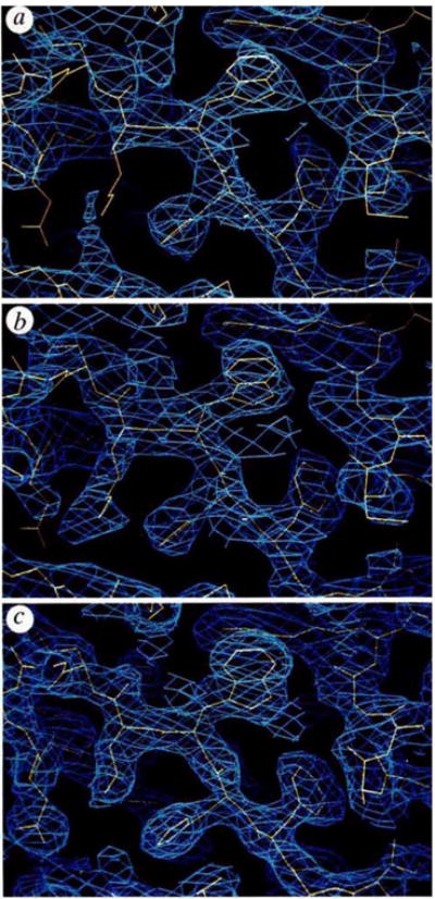
Electron-density distributions used in the structural determination. The portion displayed in each panel includes the segment Phe 26-His 27- Trp28-Lys29 with the partially refined model 5 (yellow) superimposed on the density (blue), a, Experimental map at 2.7 Å resolution based on combined MIR plus MAD phase information;
= 0.58. b, Experimental map at 2.7 Å resolution after phase refinement by solvent flattening and density truncation;
= 0.86. c, Refined 2|Fobs|−|Fcalc| map with model phases after refinement at 2.3 Å resolution; R = 0.208.
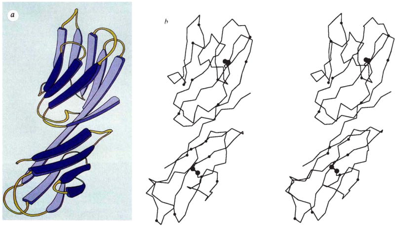
Backbone structure of the D1D2 fragment of CD4. a, Schematic diagram (copyright Yarmolinski and Hendrickson, 1990). b, Stereodiagram of the α-carbon backbone. Positions of residue numbers divisible by ten are indicated by small spheres, sulphur atoms in disulphide bridges are indicated by larger spheres. The point of view is down the Z″ axis after rotations about X(100°), Z′(−30°), Y′(−90°) and X″ (10°) starting with the frame having X, Y, Z along a, b and c*.
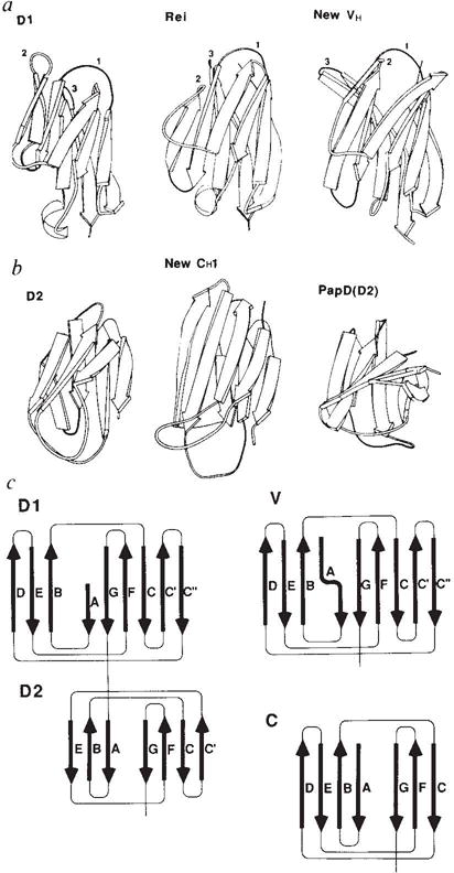
Schematic comparisons between domains of CD4 and other immunoglobulin-folded domains, a, Ribbon diagrams comparing D1 with a variable λ light-chain domain and a variable heavy-chain domain. The point of view is as in Fig. 2; the CDR loops of immunoglobulins and their analogues in D1 are indicated by numerals, b, Ribbon-diagram comparisons of D2 with a constant domain and with domain D2 of the bacterial chaperone protein PapD. c, Topology diagrams and strand nomenclature for the β sheets in CD4 and in variable and constant domains of immunoglobulins.
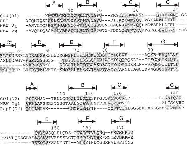
Structural alignment of the amino-acid sequences of CD4 domains with other immunoglobulin-related domains. Shaded residues have Cα positions within 2.5 Å of corresponding CD4 positions after optimal superposition of all shaded residues for a given pair of domains. (Exceptions up to 2.60 Å were allowed for residues in the middle of strands.) Each superposition relates a certain number of positions, N, within the specified 2.5 Å limit, and these match at a certain r.m.s. discrepancy, Δ. For the match of D1 with Rei (VLκ), N = 72 and Δ = 1.22 Å; for D1 versus New (VLλ), N = 66 and Δ = 1.12 Å; and for D1 versus. New (VH), N = 68 and Δ = 1.13 Å. For the match of D2 with New (CH1), N = 33 and Δ = 1.59 Å; for D2 versus PapD(D2), N = 32 and Δ = 1.26 Å; for D2 versus PapD(D1), N = 39 and Δ = 1.55 Å; and for D2 versus Rei (VLκ), N = 30 and Δ = 1.68 Å. The alignments of D2 versus PapD(D1) and D2 versus Rei (VLκ) are not shown.
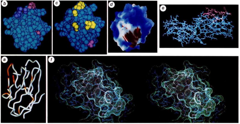
Images of CD4 relating to sites of interaction with HIV. a, Cα backbone of D1 with sites implicated by mutational and structural analysis to affect high-affinity gp120 binding (see Table 2). Non-buried residues that show reduction in binding without global disruption of structure (as judged by antibody binding) are coloured red-orange (29, 41, 42, 43, 44, 45, 47, 49, 52, 58, 59, 77, 81 and 85) on a backbone drawn by WORM (L. Andrews). The orientation is as in Figs 2 and 3. b, Van der Waals’ surface of D1 with side chains of gp120-sensitive residues coloured. Those showing marked reductions are in pink (43, 44, 85, are visible) and those showing moderate effects are in purple (59, 42, are visible). The point of view is from above a, looking down at the tips of CDR-like loops, c, Van der Waals’ surface of D1 with side chains of antibody epitopes coloured. Residues identified with epitopes of the Leu3a family and shown in purple (24, 25, 27, 42, 43, visible) and those identified with the L71 family are shown in yellow (88, 89, visible). The view is roughly as in b. d, Electrostatic potential surface computed with DELPHI at neutral pH, and displayed with AAK (A. Nicholls and B. Honig) at the levels of the solvent-accessible surface with a probe radius of 1.4 Å. Blue represents positive potential, red negative, and white neutral. The negative patch is associated with residues 85, 87 and 88. The view is approximately as in b. e, An all-atom representation of D1D2. Residues in the gp120-binding region from residue 41 to 59 are drawn in red. The direction of view is roughly as in Fig. 2, but the molecule has been rotated by ~90° about this view axis. This view illustrates the exposed nature of Phe 43. The actual conformation of this phenyl side chain in the crystal is at least partly determined by lattice interactions, but its highly exposed nature would also be expected to persist in other conformations accessible in solution, f, Stereoview of the molecular surface in the major gp120-binding region of D1. Atoms are drawn as in e, and are enveloped by the surface in contact with a probe sphere of 1.4 Å radius as displayed by QUANTA (Polygen). The view is taken after rotation from e by ~90° about the horizontal axis.
Comment in
-
Immunology. One hand clapping.Nature. 1990 Nov 29;348(6300):393-4. doi: 10.1038/348393a0. Nature. 1990. PMID: 2247144 No abstract available.
References
Publication types
MeSH terms
Substances
Grants and funding
LinkOut - more resources
Full Text Sources
Other Literature Sources
Molecular Biology Databases
Research Materials
